Please enter url.
Login
Logout
Please enter url.
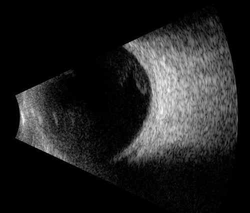
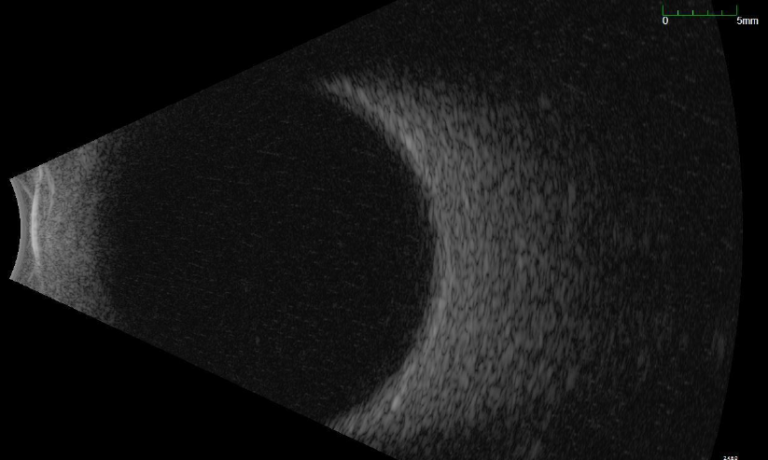
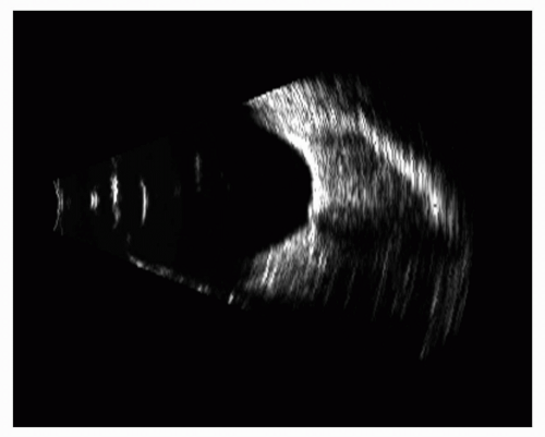
Scanmate B - The Ultra-Portable and Advanced B-scan
dghtechnology.com
source
Comments
Imaging and Treatment | Capital Eye Specialists
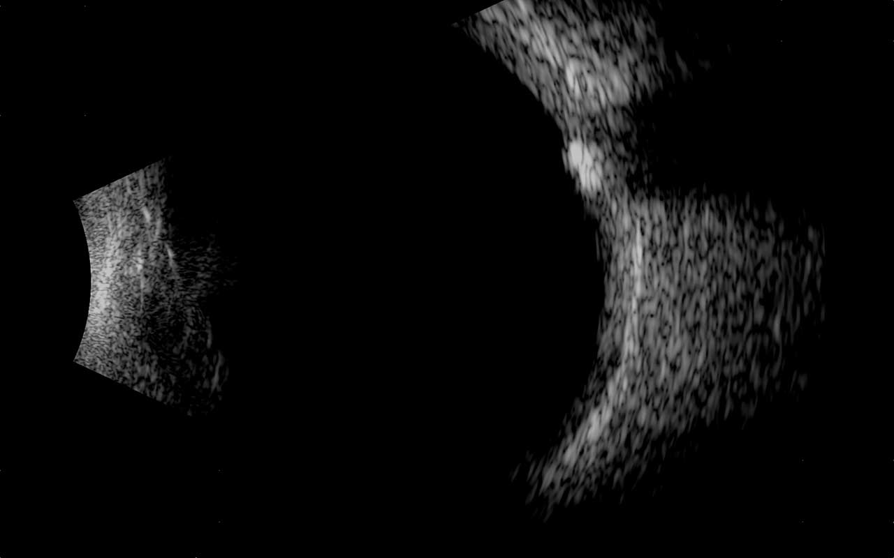
Ultrasonography of the optic nerve head: optic disc drusen. | Download ...
Ultrasound image of the left eye showing no significant vitreous ...
Scanmate B - The Ultra-Portable and Advanced B-scan
Scanmate B - The Ultra-Portable and Advanced B-scan
Nasal, longitudinal section with image suggestive of temporal mass ...
Izquierda. Engrosamiento escleral en la ecografía en modo B en ojo ...
One-Year Results of Intrastromal Corneal Ring Segment Implantation ...
Semi-Autologous Corneal Transplantation with Simultaneous Bilateral ...
Posterior vitreous detachment (PVD) grading based on B-scan ...
Symblepharon as the Only External Sign of an Occult Intraocular ...
D31.31-32 Benign Neoplasm of Choroid - Decision-Maker PLUS
Review of Retinoblastoma - American Academy of Ophthalmology
(PDF) Ultrasonographic documentation of a spontaneously resolved ...
The Mandelbrot Set has a third dimension: the Bifurcation Diagram
A and B are the anterior segment SD-OCT vertical scan of the extreme ...
B-scan ultrasound of the right eye at initial presentation shows mildly ...
Different human oocyte morphological abnormalities (arrows) observed by ...
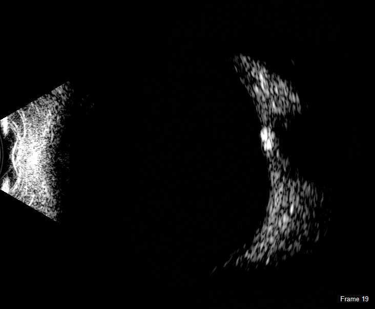
Optic nerve B-scan ultrasound in a child in which the optic nerve head ...
Spectral domain optical coherence tomography showing diabetic macular ...
Orbital Diagnosis | Radiology Key
Ocular Ultrasonography – Macula Retina Vitreous Center
Anterior segment OCT of a fully detached DMEK graft lying on the iris ...
Ecografía del ojo izquierdo, cortes axial y longitudinal. | Download ...
Case #17: Ultrasound of Intrinsic Vascularity » New York Eye Cancer Center
Control ultrasound at one year. temporal inferior longitudinal section ...
Figure 2 from Bilateral optic neuritis in pregnancy. | Semantic Scholar
Optical coherence tomography findings of falciform retinal detachment ...
B-scan ultrasound images displayed the "table top" shaped tractional ...
Figure 2 from How a Devastating Case of Acanthamoeba Sclerokeratitis ...
B-scan ultrasonography of the right eye (OD) demonstrating a single ...
Orbital Diagnosis | Radiology Key
A case of posterior ciliary artery occlusion following pneum ...
Images taken at different scales. A zoom on a particular region is ...
The Monster Black hole TON 618 : spaceengine | Black hole, Black hole ...
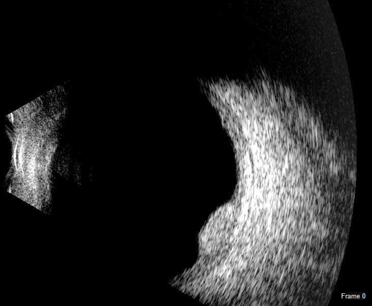
Optic-Nerve-Scan
Buried-Optic-Nerve-Drusen
Optic-Nerve-Drusen-Visual-Field
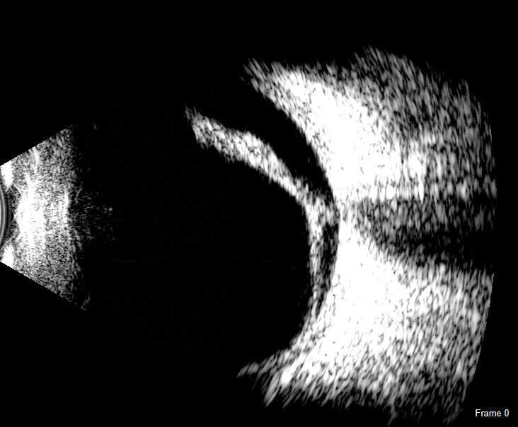
Optic-Disc-Drusen-B-scan
Optic-Nerve-Head-Drusen
Optic-Nerve-Head-Drusen-On-Oct
Optic-Nerve-Drusen-Ultrasound
Optic-Nerve-Drusen-vs-Papilledema
Optic-Nerve-Drusen-Autofluorescence
Onh-Drusen
Onh-Edema
Anterior-Ischemic-Optic-Neuropathy
Optic-Nerve-Drusen-Eye
Anomalous-Optic-Nerve
Optic-Nerve-Calcification
Optic-Nerve-Drusen-CT