Please enter url.
Login
Logout
Please enter url.
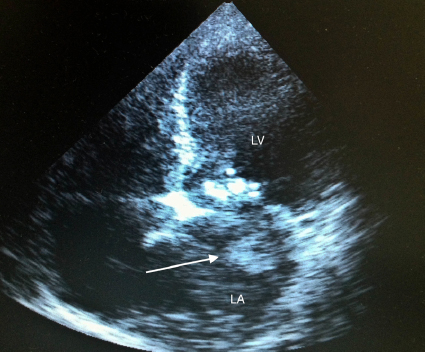
Trans thoracic echo showing the dilated right coronary artery arising ...
researchgate.net
source
Comments
Longitudinal transoesophageal view of the Cardioseal™ device with one ...
Echocardiogram demonstrating the presence of vegetation in the ...
Ecocardiograma transtorácico | Download Scientific Diagram
(a) Transthoracic echocardiogram with right ventricular myxoma (arrow ...
(PDF) Role of echocardiography in diagnosis and management of complete ...
(PDF) Acute slit-rupture of LV (“cracked heart”) complicating acute MI ...
Measurements of aortic valve complex and of the ascending aorta ...
Transesophageal short-axis view of the aorta (Ao) with the aneurysm ...
Left Ventriculogram showing apical ballooning. | Download Scientific ...
Sagittal cardiac magnetic resonance image showing the pulmonary trunk ...
9 Huge left ventricular cavity thrombus in apical twochamber ...
(PDF) Right Ventricular Rupture Caused by Prolonged Cardiopulmonary ...
Transesophageal echocardiographic image of interatrial septum's ...
Cardiologie veterinara : RIGHT HEART DISEASES IN DOG
Apical 4-chamber view demonstrating increased wall thickness of the ...
Mosaic image of blood flow passing through the muscular ventricular ...
Two-dimensional transthoracic echocardiogram in apical 4-chamber view ...
(PDF) Prominent Eustachian Valve Mimicking Thrombus in Right Atrium ...
Admission electrocardiogram showing sinus rhythm and ST-segment ...
Echocardiogram (apical four chamber view) showing dilated right ...
Contrast-enhanced transthoracic echocardiography. Passage of ...
(PDF) Haemodialysis catheter causing internal jugular vein thrombosis ...
MRI (STIR sequence) showing hyperintense signal from L5 and S1 ...
Chapter 16 - Echocardiography Board Review: 500 Multiple Choice ...
Early detection of systemic embolism: usefulness of bedside ...
Cureus | An Unusual Cause of Recurrent Syncope: Sinus of Valsalva Aneurysm
Patient's TTE before HBOT: microbubbles (Arrow) in the right ventricle ...
Perioperative Echocardiographic Features of Total Anomalous Pulmonary ...
A chest X-ray shows right atrial enlargement. | Download Scientific Diagram
Purulent fluid demonstrated within the pericardial cavity | Download ...
Transthoracic echocardiography. | Download Scientific Diagram
Apical two-chamber transthoracic echocardiogram view shows the mass in ...
Fenestration procedure and the fenestrated ASD device. | Download ...
Current concepts in mitral valve prolapse—Diagnosis and management ...
TEE view of thrombus in the left atrium attached to the pin of the ...
Coronary-Artery-Disease-Echo
Kawasaki-Coronary-Artery-Aneurysm
Coronary-Artery-Distribution-Echo
Kawasaki-MRA-Coronary-Artery
Right-Coronary-Artery-Echo
Kawasaki-Coronary-Dilalation-On-Echo
Coronary-Irregular-Kawasaki-Echo
Coronary-Echo-Hyperechogenecity-Kawasaki
Echo-Coronary-Arteries-Kawasaki
Coronary-Artery-Disease-Echo-Findings
Coronary-Dilalation-On-Echo-Kawasaki-Dis-Eas
Coronary-Artery-Distribution-EKG
Kawasaki-MRI-Coronary-Artery
Coronary-Artery-Dilatation-in-Kawasaki-Disease
High-Right-Coronary-Artery-Echo
Kawasaki-Disease-Coronary-Artery-Aneurysm-Mechanism