Please enter url.
Login
Logout
Please enter url.
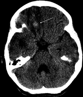
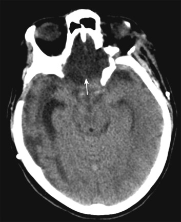
Non-contrast-enhanced axial CT image showing a hyperdense left anterior ...
researchgate.net
source
Comments
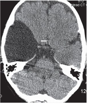
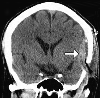
Non-contrast-enhanced axial CT image showing a hyperdense right middle ...
Axial noncontrast CT Head (a) and postcontrast T1 (b) MR images at the ...
61-year-old female with ruptured distal anterior inferior cerebellar ...
Head and Spine Trauma | Musculoskeletal Key
Hyperdense middle cerebral artery sign | Image | Radiopaedia.org
MCA Territory
ACUTE HEADACHE IN THE EMERGENCY DEPARTMENT | Journal of Neurology ...
CT venogram in intracranial venous thrombosis. Note the absence of ...
Article Fulle Text
(PDF) Hyperdense middle and anterior cerebral arteries: Familiar and ...
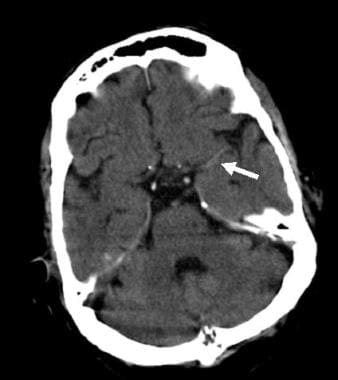
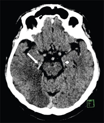
-CT of the head revealed a hyper-dense sign in the right middle ...
Isn't that CT Enough? - Water Cooler Breakdown of CT vs CT/LP for SAH ...
Computed tomography scan of the brain showing pneumocephalus in the ...
(PDF) Case 14694 Severe hypoxic-ischemic brain injury: the reversal ...
205 | Radiology Key
Cerebrovascular disease and non-traumatic haemorrhage | Radiology Key
(PDF) Acute respiratory failure after endoscopic third ventriculostomy ...
Cerebral venous thrombosis in a young female patient with ...
Imaging of initial ischemic stroke. (a) CT without contrast, showing ...
(PDF) Case 14694 Severe hypoxic-ischemic brain injury: the reversal ...
On esophagogastroduodenostopy, (A) grade III varices with red wale sign ...
Imaging Evaluation of the Adult Presenting With New-Onset Seizure | AJR
Stroke Imaging: Practice Essentials, Computed Tomography, Magnetic ...
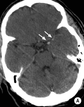
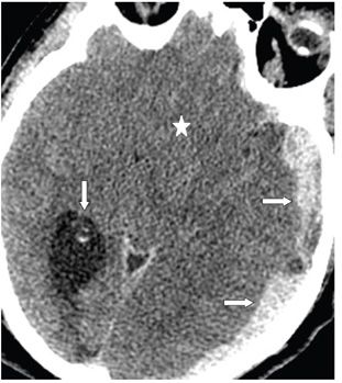
A case of large right MCA stroke with hyperdense MCA sign in CT imaging ...
Imaging of Vascular and Endovascular Surgery | Radiology Key
CT scan of the head without contrast showing diffuse encephalomalacia ...
The porencephalic cyst directly communicates with the posterior horn of ...
Axial GRE image showing extra-axial abnormal lesion(M) in right CP ...
Traumatic Brain Injury | Radiology Key
The hyperdense vessel sign in cerebral computed tomography: pearls and ...
Hyperintense lesions mainly in posterior parieto-occipital area on both ...
Coronal contrast-enhanced head CT shows mass-like thickening and ...
Three black clots retrieved from the left MCA. MCA: middle cerebral ...
207 | Radiology Key
Primary intraosseous meningioma of the calvaria | Eurorad
Hyperdense-Artery-Sign
Middle-Cerebral-Artery-Occlusion
M1-Segment-of-Middle-Cerebral-Artery
Middle-Cerebral-Artery-Territory
Middle-Cerebral-Artery-Stroke-CT
Middle-Cerebral-Artery-Segments-Anatomy
Right-Middle-Cerebral-Artery-Stroke
Middle-Cerebral-Artery-Branches-Anatomy
Dense-Artery-Sign
Middle-Cerebral-Artery-Stroke-Symptoms
Left-Middle-Cerebral-Artery-Stroke
Right-Anterior-Cerebral-Artery
Middle-Cerebral-Artery-Segments-Radiology
Hyperdense-Basilar-Artery-Sign
MCA-Infarct
Pca-Posterior-Cerebral-Artery