Please enter url.
Login
Logout
Please enter url.
(PDF) Surgical ventricular reconstruction for ischaemic heart failure ...
researchgate.net
source
Comments
Localized left ventricular remodelling involving the basal segment of ...
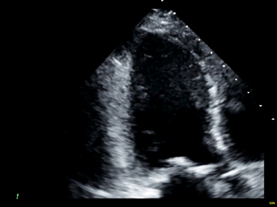
Cardiac MRI Revealing Low Ejection Fraction with Dilated Cardiac ...
(PDF) Surgical ventricular reconstruction for ischaemic heart failure ...
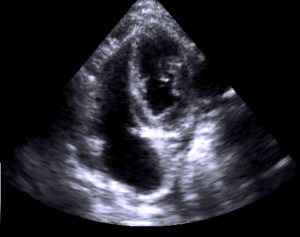
Apical four chamber view of a patient with confirmed tuberculous ...
POCUS Made Easy: eFAST • LITFL • Ultrasound Library
2D Echo showing left ventricular pseudoaneurysm (star sign). | Download ...
Incidência apical duas câmaras na telediástole que mostra a hipertrofia ...
Echocardiography demonstrating (red arrow) a large pericardial effusion ...
(a) Transesophageal echocardiogram showing homogeneously thickened ...
Gross pathological specimen of pericardium after pericardiectomy ...
Echocardiography showing septal hypertrophy in a patient with HCM ...
Septal bulge and LVOT obstruction - YouTube
Mitral valve anatomy. This figure shows 'Volume rendered images' (VRI ...
Echo Case: Right Test, Wrong Reason
Small Cell Lung Cancer Presenting as a Cardiac Mass with Embolic ...
Chest X ray (PA) showing 2 atrial and 2 ventricular pacing leads and 1 ...
Platypnea-orthodeoxia syndrome in a patient with an ascending aorta ...
A very large circumferential pericardial effusion visualized via ...
Cat restrictive cardiomyopathy (RCM)? non classified ? - Members
Basal location of LVFT. LVFT: Left ventricular false tendon | Download ...
Echocardiogram with four-chamber view before PAH therapy. | Download ...
Agitated saline test during a transthoracic echocardiogram. A positive ...
Takotsubo’s Paradox
Follow-up transthoracic echocardiography showing a scanty amount of ...
Final TEE image. Note the current septal position, indicating improved ...
Ecocardiograma transtorácico. | Download Scientific Diagram
A trans-esophageal echocardiogram showed a bioprosthetic valve in the ...
Chapter 15 - Echocardiography Board Review: 500 Multiple Choice ...
Echocardiogram - Cardiologist Dr Paul Ong | Heart Clinic
(a) PET/CT scan of the patient demonstrates normal cardiac muscular ...
Eisenmenger patient in case 1. Transthoracic echocardiogram ...
| Subxiphoid abdominal short axis view ("Situs view"). | Download ...
Apical 2 chamber view with agitated saline captured at end systole ...
Ecocardiograma con amiloidosis cardiaca - YouTube
Transoesophageal echocardiogram revealing a large posterior mitral ...