Please enter url.
Login
Logout
Please enter url.
MRI in CLN2 disease patients: Subtle features that support an early ...
ejpn-journal.com
source
Comments
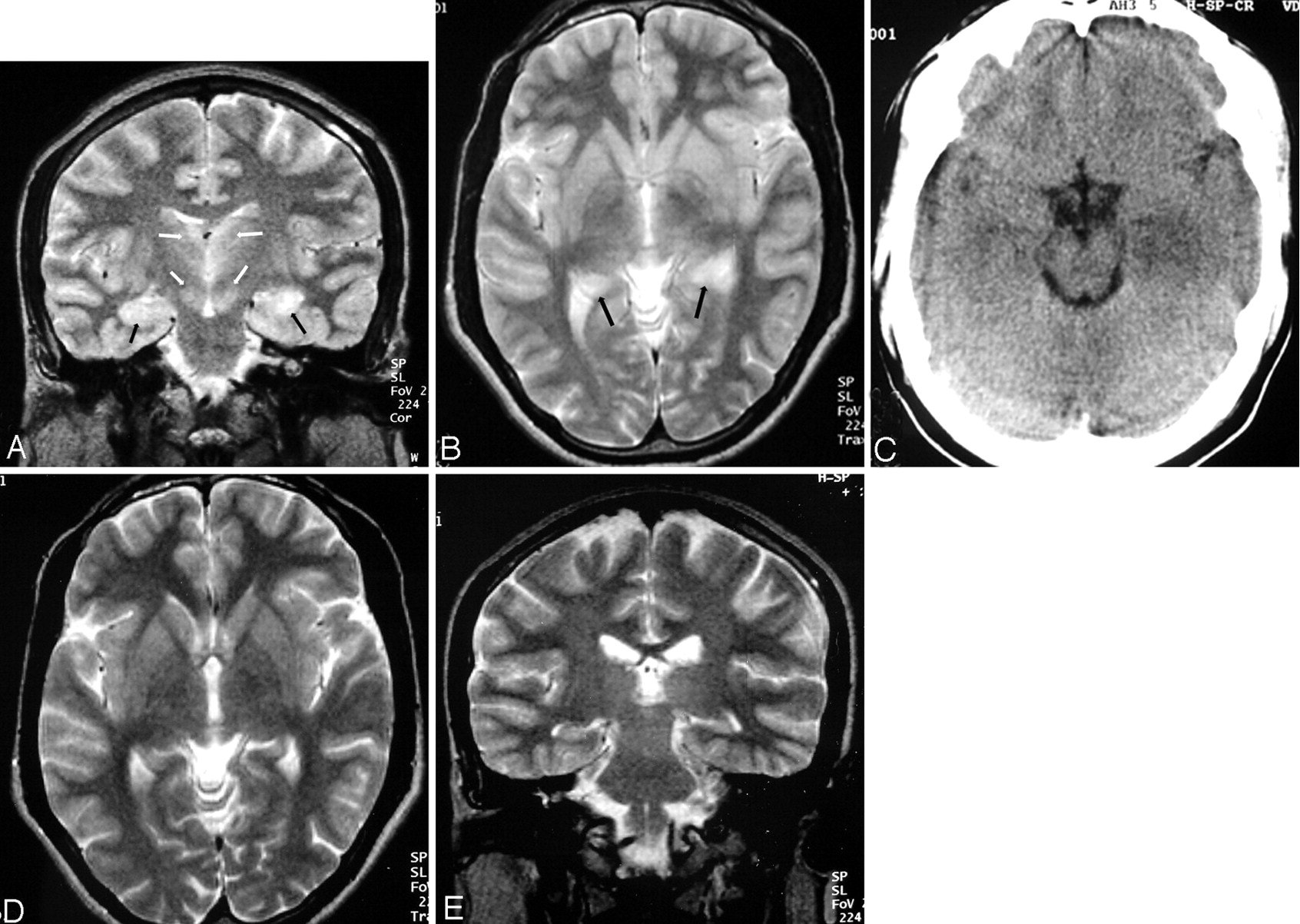
A Patient 1 e T2-weighted axial sections from an MRI scan taken 1 year ...
Intraventricular Hemorrhage - Cytotoxic Edema - Mussen Healthcare
This figure illustrates a selection of MRI features that were shown to ...
Rasmussen’s encephalitis in a 10-year old girl, a–c. Axial T2-weighted ...
Case 32-2019: A 70-Year-Old Woman with Rapidly Progressive Ataxia | NEJM
Temporal Lobe Involvement in Japanese Encephalitis: Problems in ...
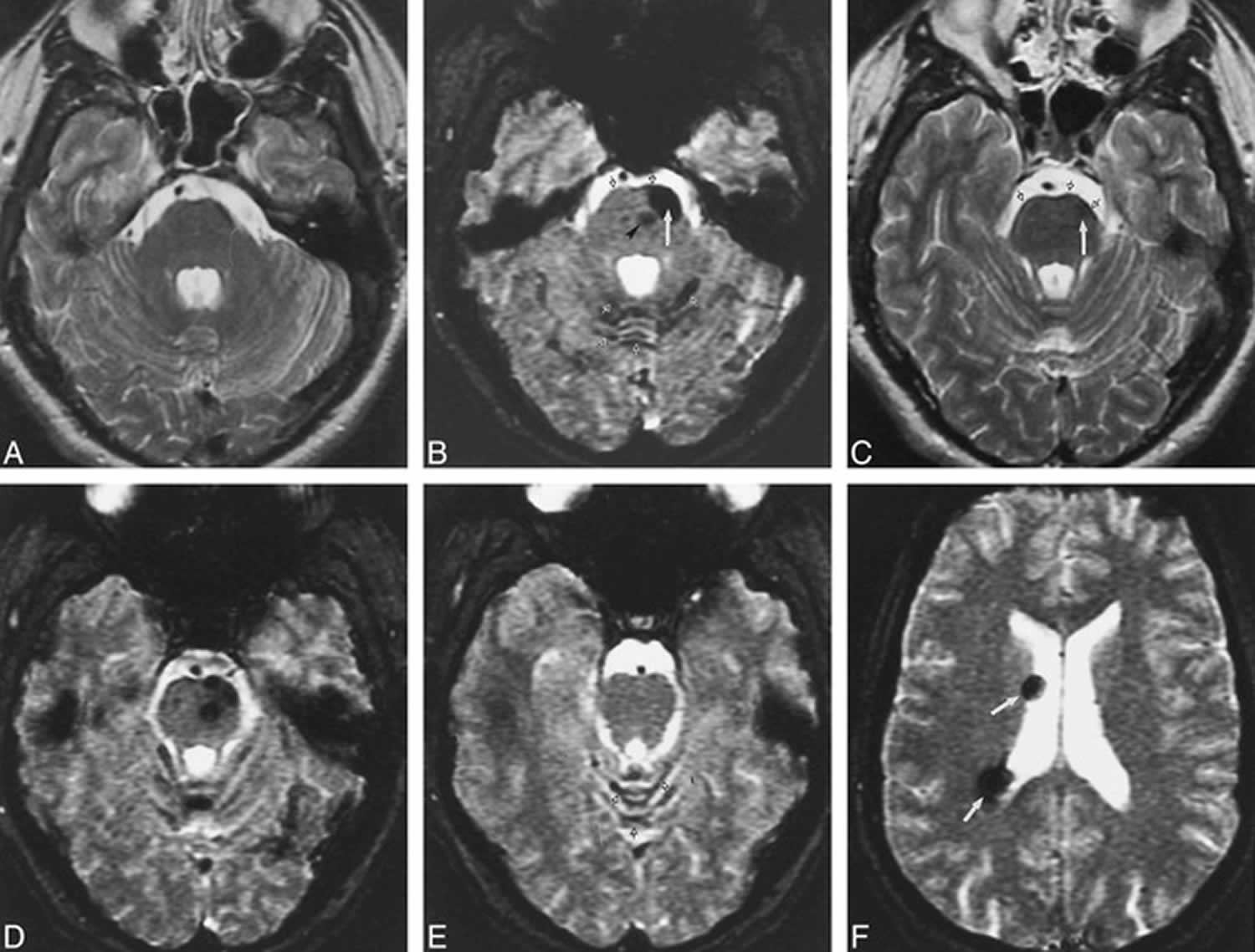
Diffusion-weighted imaging hyperintense lesions with apparent diffusion ...
Chimeric Antigen Receptor T-Cell Therapy and Imaging Applications for ...
CNS MR and CT Findings Associated with a Clinical Presentation of ...
Axial T2 (A–C) and FLAIR (D–F) showing swelling and abnormal high ...
Brain MRI of case 2 at age 57 (A-C) and 60 (D-F) showing some degree of ...
Figure 1 from Transient Lesion in the Splenium of the Corpus Callosum ...
Ophthalmic features of PLA2G6-related paediatric neurodegeneration with ...
Reversible encephalopathy caused by an inborn error of cobalamin ...
A common pattern of brain MRI imaging in mitochondrial diseases with ...
Multiple Myeloma Invasion of the Central Nervous System | Allergy and ...
Brain MRI made at the age of 13 months (images A, B, and C) and at the ...
Headache, dizziness, sensorineural hearing loss, and bilateral leg ...
e (a,b): Cortico subcortical hyperintensity in temporoparietal ...
Lymphomatosis cerebri: a treatable cause of rapidly progressive ...
Visual and neurologic sequelae of methanol poisoning in Saudi Arabia ...
Case 32-2019: A 70-Year-Old Woman with Rapidly Progressive Ataxia | New ...
Superficial siderosis definition, causes, symptoms, diagnosis ...
Neuroimaging in MIRAS disease (POLG mutation) and PENDRED syndrome ...
Serial changes in brain T2-weighted MRI findings in a patient with ...
Brain magnetic resonance (MR) images of the patient before and after ...
A 5-year-old girl with left cerebellar JPA. Residual tumor failed to ...
(a) Eleven-year-old male. T2W axial demonstrates numerous hypointense ...
Case 27-2022: A 32-Year-Old Man with Confusion, Headache, and Fever | NEJM
Axial T2 weighted images show high signal in posterior limb of internal ...
Brain MRI imaging. T2-weighted TSE axial scans (1.5 T) at the level of ...
Case 38-2017 — A 20-Year-Old Woman with Seizures and Progressive ...
Figure 1 from An unusual case of hypertensive encephalopathy involving ...
Frontiers | Clinical, radiological, and genetic characterization of ...
Occasional seizures, epilepsy, and inborn errors of metabolism - The ...