Please enter url.
Login
Logout
Please enter url.
Femoral Artery And Vein Anatomy
mungfali.com
source
Comments
(PDF) The effects of hip abduction with external rotation and reverse ...
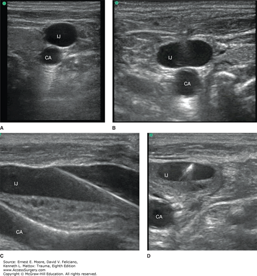
(A, B) Ultrasound images of the rIJV and relationship with rCA. (A ...
Staging of DDH according to Graf: A. Graf 2A; B. Graf 2B; C. Graf 2C ...
Ultrasound image for measuring (a) cross-sectional area (CSA) and (b ...
Standard coronal ultrasound section through the acetabulum of a normal...
(PDF) The Effectiveness Of Trendelenburg Positioning On The Cross ...
US of the Pediatric Female Pelvis | Radiology
Femoral Hernia - Risk Factors - Clinical Features - Management ...
A prospective, assessor-blind evaluation of surgeon-performed ...
Diagnostic Imaging in Patient with Elbow Pain: Findings - The ...
Table Developed to Predict Gestational Age in Quarter Horses – The Horse
A AND B: Hyperechoic fatty hilum in a cervical lymph node (left panel ...
Cross-sectional Diameters of the Internal Jugular Veins. | Download ...
Microscopic image of the tumour of the left parotid gland (a, b), right ...
Sonogram of the caudal cervical esophagus (arrows). In this horse, the ...
Calculating the tracheal index to evaluate thyroid size. Transverse ...
(PDF) Incidental subclavian artery injury during right internal jugular ...
Left lateral decubitus fluoroscopic image from conventional ...
Long-axis ultrasound image of the internal jugular vein and the ...
A AND B: Hyperechoic fatty hilum in a cervical lymph node (left panel ...
Reproductive disorders | Veterian Key
Examples of minimally complicated cysts: (A) cyst with echogenic ...
Ultrasound anatomy (left) and scheme (right) of the right phrenic nerve ...
Ultrasound finding of benign cystic nodule with lateral lymph node ...
m_moore3_ch16_f022.png | Abdominal Key
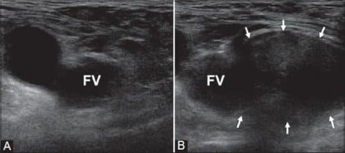
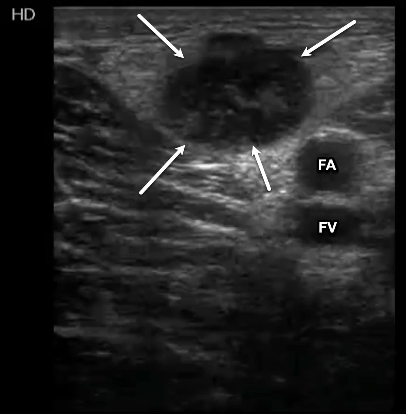
POCUS Spotlight: Lower Extremity DVT Scanning
B: Annotated ultrasound image (FA: femoral artery; FN: femoral nerve ...
The Hip - TeachMe Orthopedics
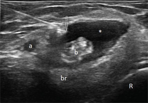
Moving below the inguinal ligament, the femoral artery and vein are ...
(PDF) [Basic physical principles of ultrasonography, anatomy of the ...
e MR imaging of the nerve sheath tumor (sagittal and coronal section ...
Case 290 | Radiology
Normal appendix. a, b Transverse gray-scale US images with (a) and ...
Distribución del anestésico local en el abordaje infraclavicular, PM ...
SciELO - Brasil - Cysts within Otherwise Probably Benign Solid Breast ...
Femoral-Vein-On-Ultrasound
Femoral-Artery-Ultrasound
Femoral-Nerve-Artery/Vein
Deep-Femoral-Vein-Ultrasound
Common-Femoral-Artery-and-Vein
Ultrasound-Image-of-Superficial-Femoral-Vein-and-Superficial-Femoral-Artery
Basilic-Vein-Ultrasound
Femoral-Artery-Adductor-Canal
Greater-Saphenous-Vein-Ultrasound
Ultrasound-Visualization-of-Femoral-Artery
Femoral-Artery-Pseudoaneurysm-Ultrasound
Femoral-Artery-Access
Femoral-Vein-Anatomy-Ultrasound
Femoral-Vein-Catheter
Common-Iliac-Vein-Ultrasound
Popliteal-Vein-Ultrasound