Please enter url.
Login
Logout
Please enter url.
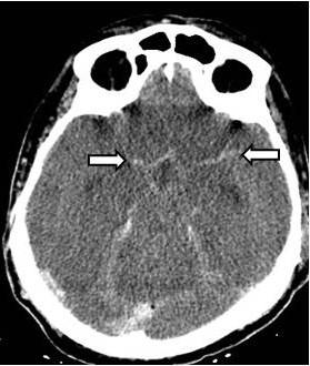
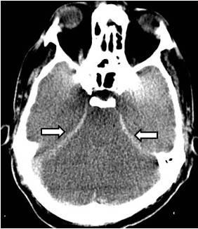
Severe hypoxic - ischaemic brain injury: the reversal, the pseudo ...
eurorad.org
source
Comments
Severe hypoxic - ischaemic brain injury: the reversal, the pseudo ...
Severe hypoxic - ischaemic brain injury: the reversal, the pseudo ...
(PDF) Case 14694 Severe hypoxic-ischemic brain injury: the reversal ...
Severe hypoxic - ischaemic brain injury: the reversal, the pseudo ...
(PDF) Case 14694 Severe hypoxic-ischemic brain injury: the reversal ...
Severe hypoxic - ischaemic brain injury: the reversal, the pseudo ...
Imaging in Acute Stroke and TIA – EmergencyPedia
Severe hypoxic - ischaemic brain injury: the reversal, the pseudo ...
CT scan showing bilateral, symmetric calcifications in thalamus, basal ...
Contrast-enhanced MDCT of neck and thorax, showing partial occlusion of ...
[Fall 2013 Block III] Skull Fractures, Traumatic Brain Injury, and ...
Sagittal section of brain CT showing significant pneumocephalus with ...
Non-aneurysmal perimesencephalic subarachnoid haemorrhage | Eurorad
Head Injury | Anesthesia Key
CT scan at admission; hyperdensity (arrow) indicates intratumoral ...
Imaging of initial ischemic stroke. (a) CT without contrast, showing ...
Stroke Imaging: Practice Essentials, Computed Tomography, Magnetic ...
Article Fulle Text
Wake-Up Call: Pulmonary Arteriovenous Malformation - The American ...
61-year-old female with ruptured distal anterior inferior cerebellar ...
Non-Communicating Hydrocephalus - The Western Journal of Emergency Medicine
Results of CT scan showing a large hyperdense right parietal convexity ...
MRI of the head revealed a large left-sided arachnoid cyst (arrows) and ...
CT image showing thickening of the posterior part of the right eyeball ...
Functional endoscopic sinus surgery (FESS): Is it always a safe procedure?
lumbar puncture Archives - Charlie's ED
Axial contrast HRCT scan showing hyperdense mass occupying the right ...
Axial NCCT scan depicts hyperdensity of right ICA distal bifurcation ...
An Elderly Woman with the GáCS Sign - Journal of Emergency Medicine
Brain computed tomography shows epidural hematoma on left temporal area ...
Meningococcal Meningitis
Brain abscess caused by Citrobacter koseri infection in an adult ...
Computed tomography scan of the brain showing pneumocephalus in the ...
RadiologySpirit: Corpus Callosal Anomalies
a) Axial MRI T2 image of the posterior fossa shows bilateral normal ...
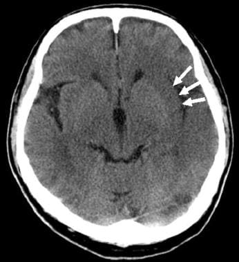
Subarachnoid-Haemorrhage-CT
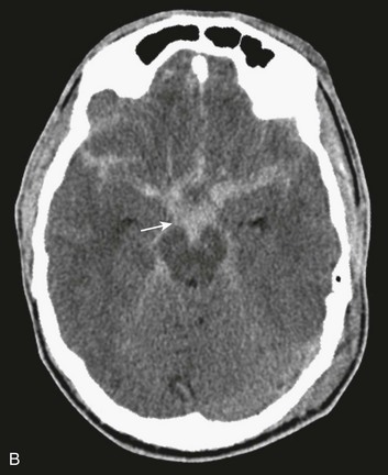
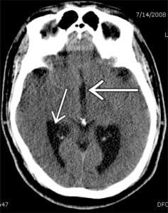
Pseudo-Subarachnoid-Hemorrhage
Subarachnoid-Hemorrhage-On-CT-Scan
Acute-Subarachnoid-Hemorrhage
Subarachnoid-Cyst-CT
Subarachnoid-Hemorrhage-MRI
Subarachnoid-Hematoma
Subarachnoid-Brain-Hemorrhage
Subarachnoid-Star-Sign
Pseudo-Sah
Subarachnoid-Hemorrhage-Treatment
Subarachnoid-Hemorrhage-Diagram
Subarachnoid-Space
Basal-Cistern-Subarachnoid-Hemorrhage
Subependymal-Hemorrhage
Perimesencephalic-Sah


![[Fall 2013 Block III] Skull Fractures, Traumatic Brain Injury, and ...](https://o.quizlet.com/Y5qzRZ.p3UKMLtn8S5Uu0g_m.png)