Please enter url.
Login
Logout
Please enter url.
The Clinical Picture: Cholesteatoma - Healthcare Communications Network
hcn.health
source
Comments
Cholesteatoma | Cleveland Clinic Journal of Medicine
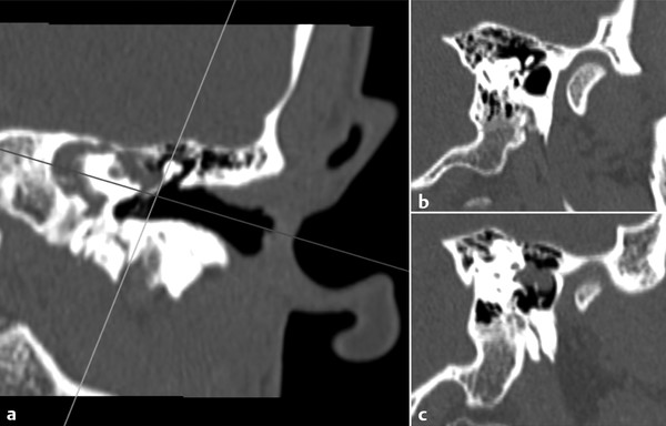
Cochlear Implantation: Systematic Approach to Preoperative Radiologic ...
The Middle Ear and Mastoid | Radiology Key
(PDF) Otosclerosis and complications of stapedectomy: CT and MRI ...
Axial and coronal CT scans showing a soft tissue mass (marked by the ...
Postoperative computed tomography of frontal recess cells. a Complete ...
Cochlear Implantation: Systematic Approach to Preoperative Radiologic ...
Imaging of Jugular Foramen | Radiology Key
Fibrous dysplasia of the temporal bone secondary to ear surgery: a case ...
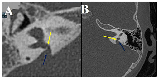
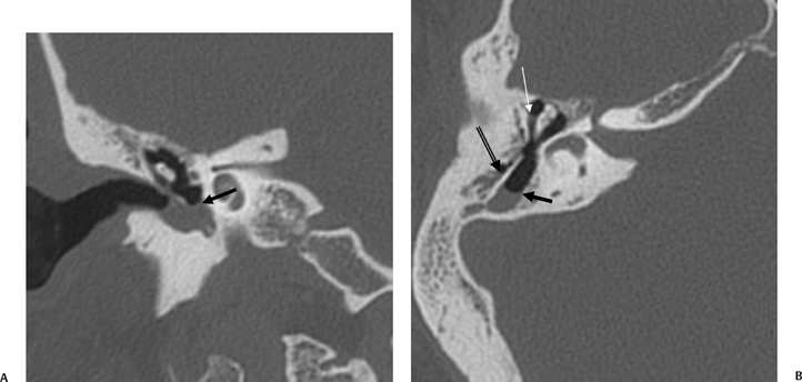
Axial contiguous CT images of left temporal bone (more superior [A] and ...
EAC cholesteatoma. Axial (a) and coronal (b) CT images show a lobulated ...
(A) Axial CT image showing intravestibular medial migration of the ...
Frontiers | Proposal for a Unitary Anatomo-Clinical and Radiological ...
Maturation of the anterior petrous apex: surgical relevance for ...
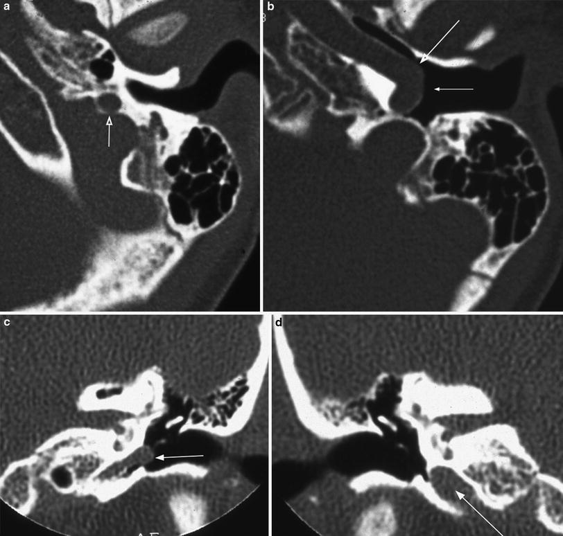
Serial axial sections of the temporal bone demonstrate complete ...
Imaging of the Postoperative Ear and Temporal Bone | Radiology Key
Chronic inflammatory middle ear disease: Postoperative CT and MRI ...
Barotrauma Presenting as Temporal Lobe Injury Secondary to Temporal ...
Non-Syndromic Sensorineural Hearing Loss in Children - Neuroimaging Clinics
The External Auditory Canal and Pinna | Radiology Key
Imaging of the Mastoid, Middle Ear, and Internal Auditory Canal After ...
Cureus | Late Recurrence of a Rare Middle Ear Neuroendocrine Tumor With ...
Endoscopic Ear Surgery | Ento Key
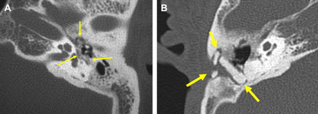
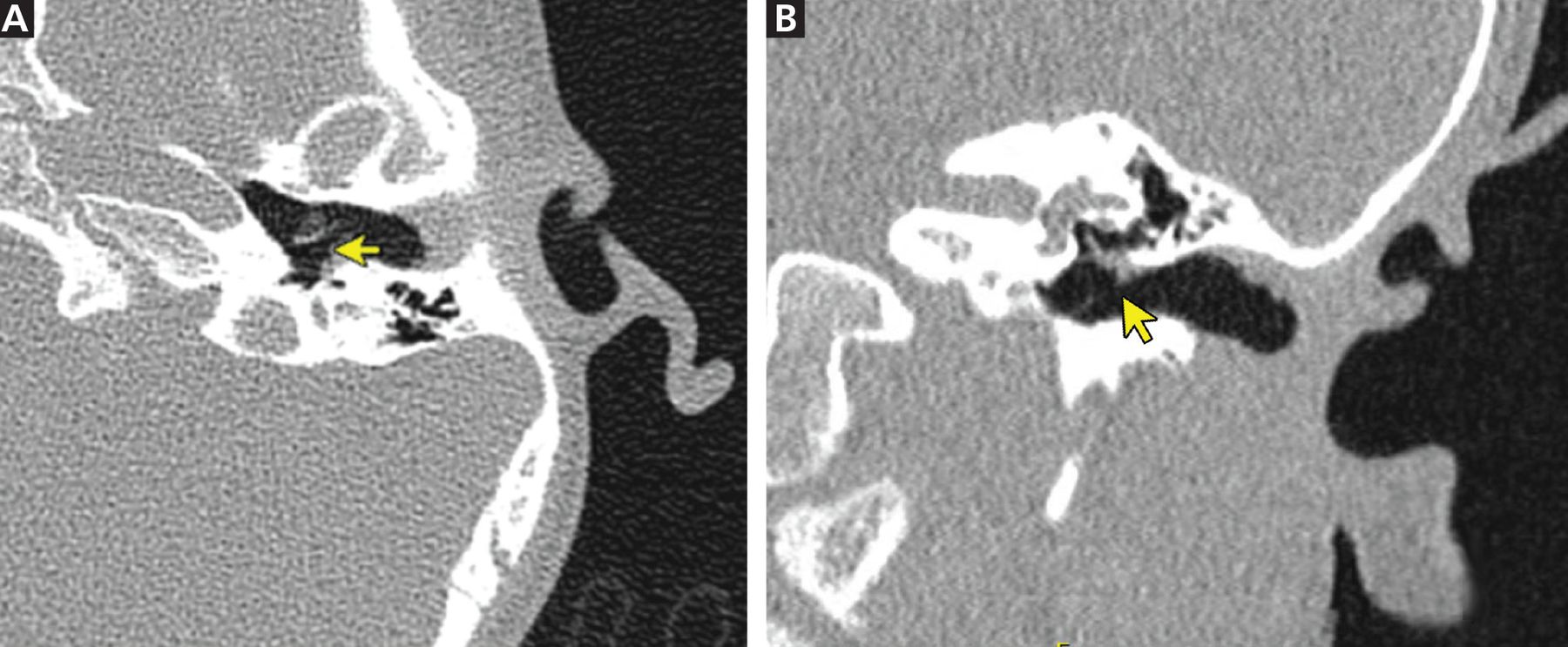
Right temporal bone CT scans in axial view showing (white arrowheads ...
Cochlear Implantation: Systematic Approach to Preoperative Radiologic ...
High-Resolution CT Findings in Children with a Normal Pinna or Grade I ...
The Middle Ear and Mastoid | Radiology Key
Congenital malformations of the external and middle ear - European ...
Congenital cholesteatoma. (a) axial section and (b) coronal section CT ...
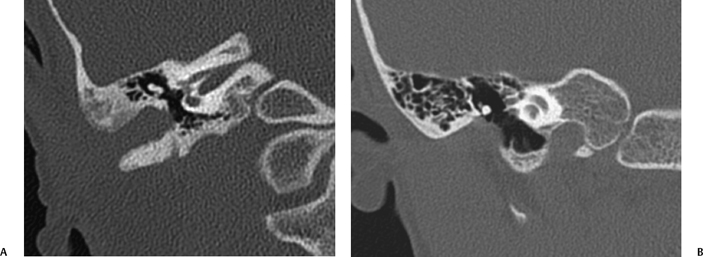
Postoperative computed tomography scans showing (A) traditional and (B ...
Temporal Bone Lesions | Radiology Key
Radiological assessment of the sinus tympani: temporal bone HRCT ...
MDCT scan shows method of VA measurement at the midpoint (black filled ...
| Left temporal bone high-resolution computed tomography (HRCT ...
Deiscenza della II porzione del faciale: in a aspetto normale della ...


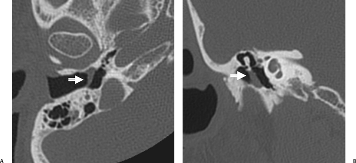


![Axial contiguous CT images of left temporal bone (more superior [A] and ...](https://www.researchgate.net/profile/Mehmet-Gencturk/publication/327142275/figure/fig11/AS:662156676722709@1534881862430/Axial-contiguous-CT-images-of-left-temporal-bone-more-superior-A-and-more-inferior.png)