Please enter url.
Login
Logout
Please enter url.
Repeated magnetic resonance imaging and cerebral performance after ...
resuscitationjournal.com
source
Comments
Repeated magnetic resonance imaging and cerebral performance after ...
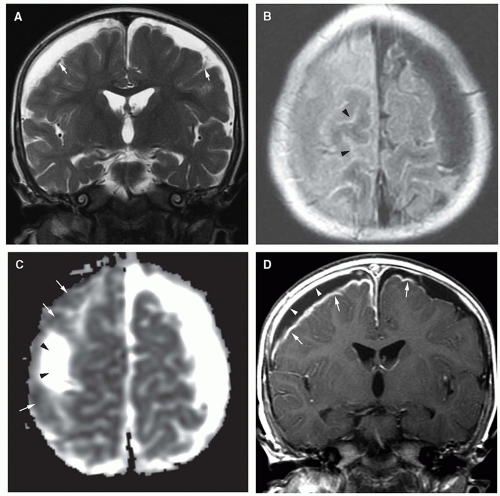
Wernicke’s encephalopathy-like lesions in global cerebral hypoxia ...
Leukoaraiosis and Stroke | Stroke
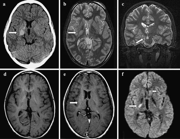
Bilateral basal ganglia lesions at presentation (a–e): hypodense on CT ...
a DWI with intratumoral bleeding in a GBM patient without AT. b ...
PMG. Axial T2-weighted image shows microgyria with normal cortical ...
Figure 1 from Creutzfeldt-Jakob Disease Presenting with Dementia and ...
Patient 1: facial features with down-slanting palpebral fissures ...
Anti-NMDAR autoimmune encephalitis - Brain and Development
Infections of the Developing and Mature Nervous System | Radiology Key
Intraocular Lymphoma: Ten Points Every Ophthalmologist Should Know
Bupropion Overdose Presenting as Status Epilepticus in an Infant ...
Chimeric Antigen Receptor T-Cell Therapy and Imaging Applications for ...
Early hyperacute stroke in a 49-year-old woman with right lower ...
The Present and the Future of Neuroimaging in Amyotrophic Lateral ...
Cerebral small vessel disease: Recent advances and future directions ...
MEFV gene mutations in neuro‐Behçet's disease and neuro‐Sweet disease ...
Magnetic resonance imaging in Case 4 show a right perisylvian mass ...
(a and b) Cranial magnetic resonance imaging showing high signal in the ...
Figure 12 from Hypoxic-ischemic brain injury: imaging findings from ...
Inherent diagnostic and treatment challenges in germinoma of the basal ...
“Thalamic aphasia” after stroke is associated with left anterior lesion ...
The protean manifestations of central nervous system IgG4-related ...
Diffusion-Weighted MRI in 300 Patients Presenting Late With Subacute ...
Visual and neurologic sequelae of methanol poisoning in Saudi Arabia ...
Cerebral MRI abnormalities associated with vigabatrin therapy - Pearl ...
MRI brain: diffusion-weighted imaging | Download Scientific Diagram
Moderate posterior reversible encephalopathy syndrome (PRES) in an ...
Imaging and CSF analyses effectively distinguish CJD from its mimics ...
Representative brain MRI findings of the patients with small vessel ...
Figure 1 from Scattered Brain Infarct Pattern on Diffusion-Weighted ...
EPOS™
The spectrum of associated brain lesions in cerebral sinovenous ...
Creutzfeldt-Jakob Disease with a prion protein gene codon 180 mutation ...
Movement disorder and neuronal migration disorder due to ARFGEF2 ...
MRI-Exam
Cardiac-CT-Scan
MRI-Patient
MRI-Test
MRI-of-Heart
MRI-Process
MRI-Views
Chest-MRI
CT-Heart-Anatomy
CT-Coronary-Angiogram
Normal-Heart-CT-Scan
Cardiac-Catheterization
EKG
Myocardial-Amyloidosis
Left-Atrium-CT
Cardiac-MRI-Labelled