Please enter url.
Login
Logout
Please enter url.
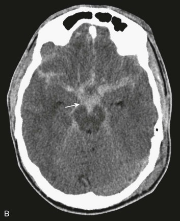
Typical CT scan showing subarachnoid haemorrhage. The image is useful ...
researchgate.net
source
Comments
Typical CT scan showing subarachnoid haemorrhage. The image is useful ...
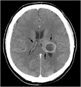
CTA brain showing PCom artery aneurysm. (a) CTA: an aneurysm arising ...
Midline shift - Wikipedia, the free encyclopedia | Vertebral artery ...
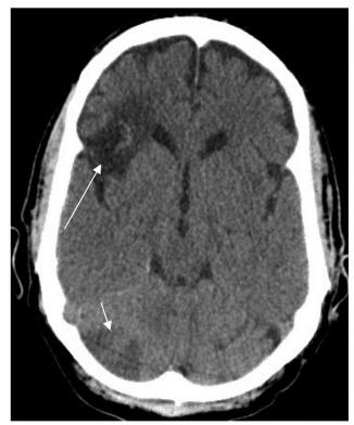
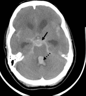
First CT scan brain without contrast showing suspicion of subarachnoid ...
Head Injury | Anesthesia Key
Acute diffuse subdural hemorrhage along the right cerebral hemisphere ...
Computed tomography angiogram scan of the head showing postoperative ...
Functional Complications: Hyperdrainage | Neupsy Key
EPOS™
EPOS™
Tumor Embolism Presenting as Rapidly-forming Cavitary Lesion | Sweigart ...
Traumatic Brain Injury Flashcards | Quizlet
(PDF) Hyperdense middle and anterior cerebral arteries: Familiar and ...
Isn't that CT Enough? - Water Cooler Breakdown of CT vs CT/LP for SAH ...
Cerebral Aneurysms and Cerebrovascular Malformations | Radiology Key
Figure. T2-weighted MRI reveals hyperintensity in basilar cistern ...
Three black clots retrieved from the left MCA. MCA: middle cerebral ...
Head Injury and Facial Trauma | Clinical Gate
The porencephalic cyst directly communicates with the posterior horn of ...
Thalamic Glioma – Toronto Notes
Neurosurgery | Basicmedical Key
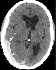
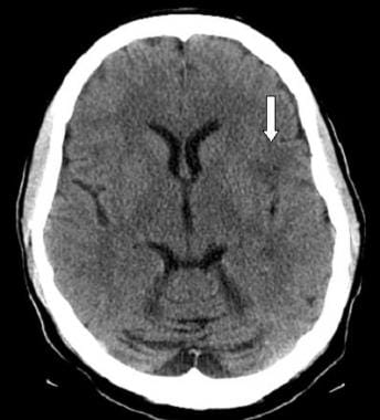
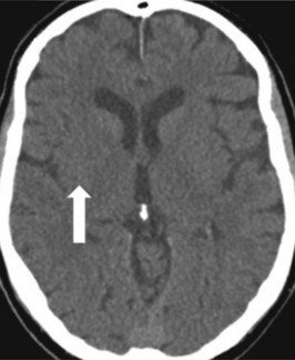
Axial CT scan of brain without contrast. Small lacunar infarct ...
Cerebrovascular Accident in a Pediatric Patient Presenting With ...
Acute subdural hematoma covering the right cerebral hemisphere (arrows ...
NeuExcell to reach the full potential of their innovative gene ...
Diffuse axonal injury. Non-enhanced CT shows multiple punctuate...
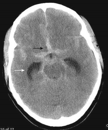
Periventricular cap hypodensity around frontal horns indicate ...
Radiology of terror injuries: the American University of Beirut Medical ...
Follow-up brain CT scan showing the newly developed ischemic area ...
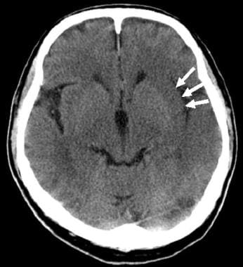
Stroke Imaging: Practice Essentials, Computed Tomography, Magnetic ...
Imaging in Acute Stroke and TIA – EmergencyPedia
Stroke Imaging: Practice Essentials, Computed Tomography, Magnetic ...
Brain Injury Education | Vanderbilt Traumatic Brain Injury Center
Brain computed tomography shows epidural hematoma on left temporal area ...
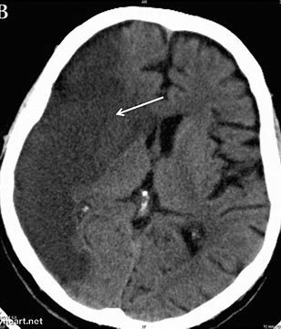
A 68-year-old woman with a left hemiplegia following a conscious ...
Subarachnoid-CT
Subarachnoid-Aneurysm
Arachnoid-Hemorrhage
Subarachnoid-Hematoma-CT
Arterial-Hemorrhage
Subarachnoid-CT-Scan
Aneurysmal-SAH
Subarachnoid-Bleed-CT
Brain-Hemorrhage-CT-Scan
Intracerebral-Hemorrhage-CT
Subarachnoid-Haemorrhage
Pseudo-Subarachnoid-Hemorrhage
Interhemispheric-Hemorrhage
Intraventricular-Hemorrhage-CT
Subarachnoid-Bleeding
Intracranial-Hemorrhage-CT-Scan