Please enter url.
Login
Logout
Please enter url.
Lecture 25: Liver & Spleen Flashcards | Quizlet
quizlet.com
source
Comments
T1 magnetic resonance angiography (MRA) of the abdomen and pelvis ...
Beata Naumnik | Professor | Medical University of Bialystok, Białystok ...
Visceral leishmaniasis causing life-threatening digestive bleeding ...
MR Imaging of Renal Masses: Correlation with Findings at Surgery and ...
Image | Radiopaedia.org
Image | Radiopaedia.org
Radiodiagnosis - Imaging is Amazing-Interesting cases: Renal vein ...
CT and MR Imaging for Evaluation of Cystic Renal Lesions and Diseases ...
Coronale reconstructie op een „contrast-enhanced computed tomography ...
Contrast-enhanced MRI of the abdomen on initial staging of the renal ...
In utero and postnatal imaging findings of parasitic conjoined twins ...
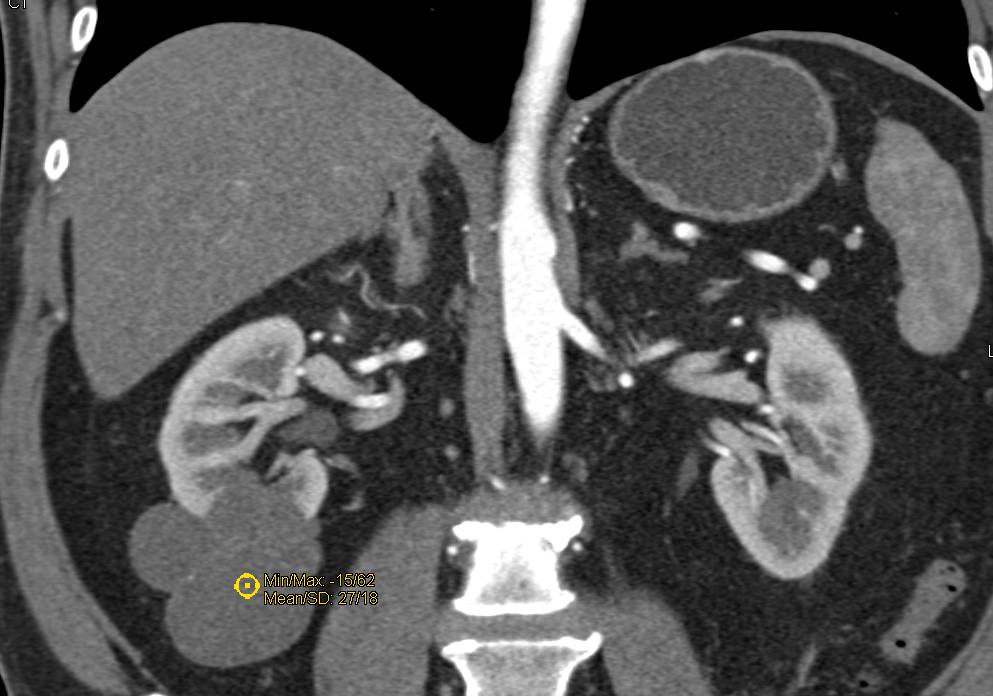
Bosniak 2 Cyst Left Kidney - Genitourinary Case Studies - CTisus CT ...
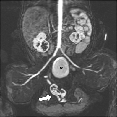
(a) Coronal 10 cm thick slab maximum intensity projection (MIP) and (b ...
Infecciones de Vías urinarias | Flowchart
RM axial en T1, que muestra riñones con pobre diferenciación ...
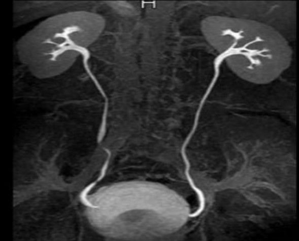
MR Urography: Techniques and Clinical Applications | RadioGraphics
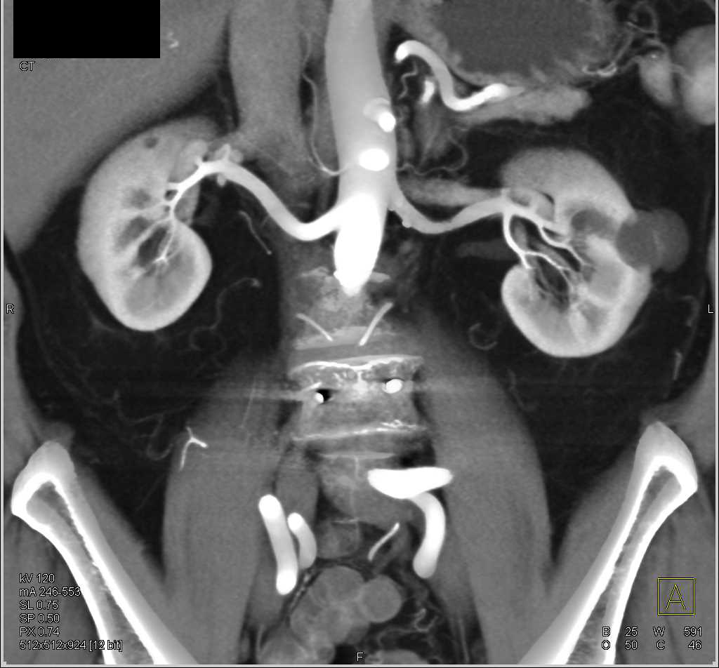
MDCT Evaluation of Ureteral Tumors: Advantages of 3D Reconstruction and ...
GU - Ureters & Bladder Flashcards | Quizlet
T2-weighted dorsal image of rabbit pelvis (at the level, close to the ...
Polypoid Mass in Left Renal Pelvis was a Transitional Cell Carcinoma ...
Spontaneous perinephric urinoma complicating obstructive uropathy | Eurorad
Bosniak II Right Renal Cyst with Thin Septations - Kidney Case Studies ...
Interruption of the inferior vena cava with aneurysm of the right renal ...
CT urogram. A maximum intensity projection, coronal CT | Open-i
Optimizing Detectability of Renal Pathology With MDCT: Protocols ...
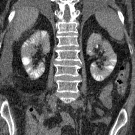
Clubbed calyces and diffuse parenchymal enhancement of the kidney ...
Fat-containing Lesions of the Retroperitoneum: Radiologic-Pathologic ...
Pediatric MR Urography: Indications, Techniques, and Approach to Review ...
What Is the Current Role of CT Urography and MR Urography in the ...
Striated nephrogram | Radiology Reference Article | Radiopaedia.org
Figure 2 from Ormond’s Disease or Secondary Retroperitoneal Fibrosis ...
Inferior vena cava (IVC) thrombectomy specimen. The left portion is the ...
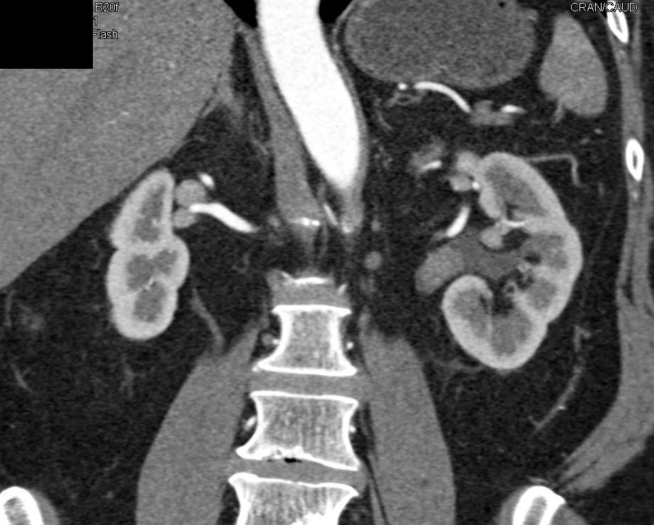
CT Urography | AJR