Please enter url.
Login
Logout
Please enter url.
Image | Radiopaedia.org
radiopaedia.org
source
Comments
Image | Radiopaedia.org
Image | Radiopaedia.org
Uterine fibroid (virtual reality) | Image | Radiopaedia.org
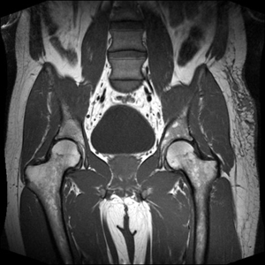
Basic-msk-mri
PLAN ARTHROGRAM HIP stir cor - mrimaster
FULL TEXT - An unusual hip dislocation during tennis playing ...
Solitary Bone Cyst | Eurorad
Basic-knee-mri
Rectus femoris avulsion injury | Radiology Case | Radiopaedia.org
Image | Radiopaedia.org
(a–c) Axial and coronal pd_tse_fs MRI pictures showing a type 1A lesion ...
Not Your Everyday Knee Pain
Image | Radiopaedia.org
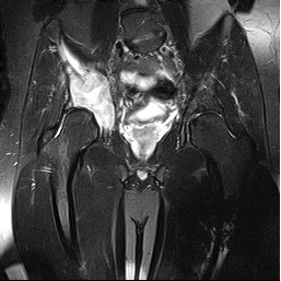
Hip Avascular Necrosis (Osteonecrosis) | AVN | Necrosis of the Femoral ...
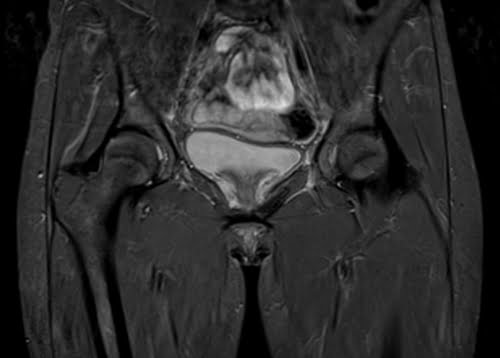
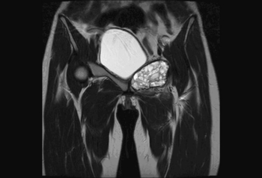
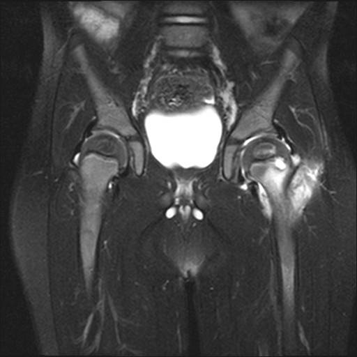
Coronal T2WI image of pelvis shows fluid -fluid levels | Open-i
Image | Radiopaedia.org
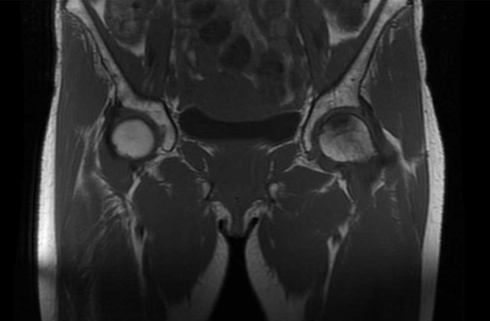
Hip joint MRI: MRI
Image | Radiopaedia.org
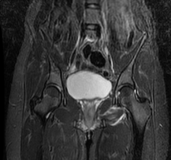
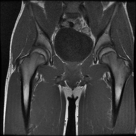
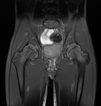
Coronal T1-weighted MR image of the pelvis. There is diffuse ...
Image | Radiopaedia.org
Image | Radiopaedia.org
Pelvic osteomyelitis | Radiology Case | Radiopaedia.org
Subchondral insufficiency fracture - femoral head | Image | Radiopaedia.org
Proximal rectus femoris tendon tear | Image | Radiopaedia.org
Slipped upper femoral epiphysis | Radiology Reference Article ...
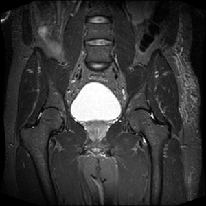
Coronal T1-weighted pelvic magnetic resonance imaging scan showing ...
Image | Radiopaedia.org
Image | Radiopaedia.org
Transient osteoporosis of the hip | Image | Radiopaedia.org
Usefulness of Dynamic Contrast-Enhanced MRI in Differentiating Between ...
Image | Radiopaedia.org
Slipped Upper Femoral Epiphysis-MRI - Sumer's Radiology Blog
Legg-Calvé-Perthes disease - biateral | Radiology Case | Radiopaedia.org
Image | Radiopaedia.org