Please enter url.
Login
Logout
Please enter url.
A) 1.5 Tesla axial diffusion-weighted MR and (B) FLAIR MR images in an ...
researchgate.net
source
Comments
MRI Based Preterm White Matter Injury Classification: The Importance of ...
Projects:RegistrationLibrary:RegLib C42 - NAMIC Wiki
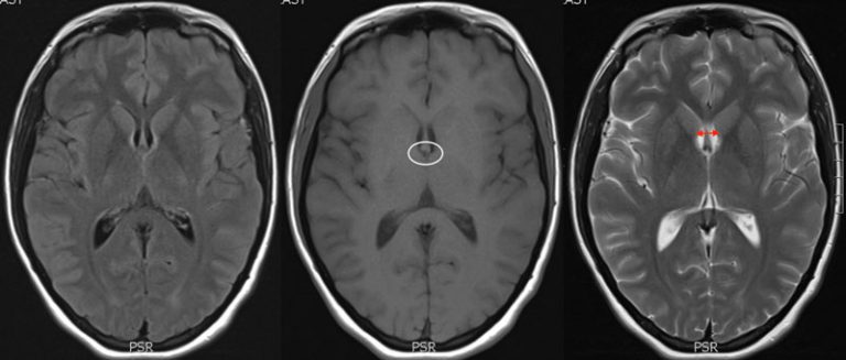
Axial T1 image, showing corresponding low signal in bilateral thalami ...
Colloid Cyst - Condition, Treatments, Recovery Times
Neuroimaging of Emergent and Reemergent Infections | RadioGraphics
Cureus | Atrial Myxoma Presenting as an Atypical Stroke in a Young ...
Parkinsonism & Related Disorders
Axial T2 weighted images showing enlargement of the subarachnoid spaces ...
(PDF) Superficial hemosiderosis of central nervous system (CNS) in ...
Eastern Equine Encephalitis
Brain magnetic resonance imaging shows diff use hypoxic brain damage ...
A Case of Narcolepsy Type 2 and Postural Tachycardia Syndrome Secondary ...
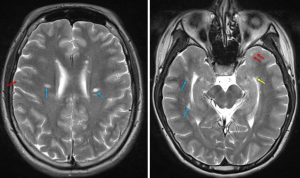
(A) T2 weighted axial MRI image showing hyperintense bilateral and ...
Interval neuroradiographic progression | Download Scientific Diagram
Paediatric non-ketotic hyperglycaemic hemichorea–hemiballismus | BMJ ...
Bilateral mesial temporal sclerosis and left temporo‐occipital cortical ...
MRI and CT appearances in metabolic encephalopathies due to systemic ...
Brain MRI Axial (A), Coronal (B) and Sagittal (C) T1-w: cerebellar ...
Brain MRI of proband 1 performed at the age of 7 years (a–c). Brain MRI ...
Dawson fingers (multiple sclerosis) – Radiology Cases
Examples of MRI scans of patients. MRI scan of one patient with ...
Coronal section of follow-up MRI, showing an ischemic stroke restricted ...
MRI brain examination of a 48-year-old male with meningoencephalitis ...
Axial contrast-enhanced T1-weighted MRI showing tuberculous meningitis ...
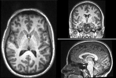
MR images demonstrating implantation of CM DBS electrodes. A ...
Projects:RegistrationLibrary:RegLib C09 - NAMIC Wiki
Brain Atrophy - Introduction
Autopsy-proved HIV encephalitis in a 35-year-old patient with AIDS ...
Magnetic resonance imaging scan of the patient's brain, including T 2 ...
Brain MRI shows multiple T2-hyperintense lesions in both supra- and ...
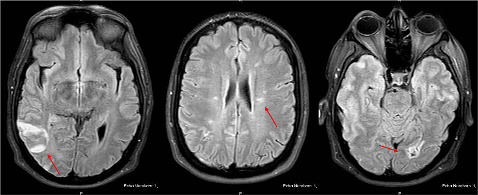
Initial magnetic resonance imaging (MRI) of the brain on axial sequence ...
coupe axiale d'une IRM cérébrale montrant un hypersignal T2-FLAIR, en ...
EPOS™
A brain CT scan of an 8-year-old boy with Hunter syndrome (MPS II ...
A 53-year-old woman with an history of chronic alcohol abuse presented ...
Cortical-Atrophy
Diffuse-Cortical-Atrophy
Diffuse-Cortical-Necrosis
Cortical-Dementia
Diffuse-Cortical-Necrosis-Kidney
Subcortical-Atrophy
Renal-Cortical-Necrosis
Global-Cortical-Atrophy
Cortical-Ischemia
Cortical-Injury
Posterior-Cortical-Atrophy
Diffuse-Cerebral-Dysfunction
Diffuse-Cortical-Laminar-Necrosis
Diffuse-Cortical-Atrophy-MRI
Acute-Cortical-Necrosis
Cortical-Mantle