Please enter url.
Login
Logout
Please enter url.
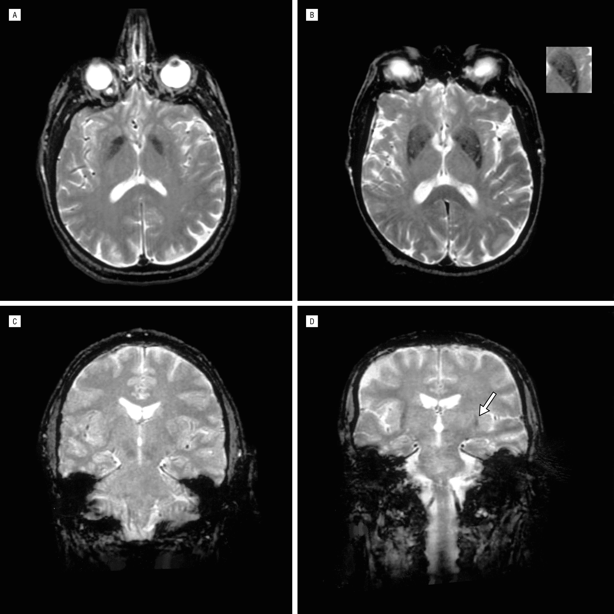
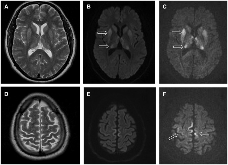
Figure 1 from [A case of vascular parkinsonism associated with ...
semanticscholar.org
source
Comments
Brain Damage by Mild Metabolic Derangements in Methylmalonic Acidemia ...
Chimeric Antigen Receptor T-Cell Therapy and Imaging Applications for ...
Hemorrhage | Neupsy Key
Visual and neurologic sequelae of methanol poisoning in Saudi Arabia ...
Parkinson's Brain Mri Vs Normal - ParkinsonsInfoClub.com
Figure 1 from Transient Lesion in the Splenium of the Corpus Callosum ...
Coinfection of Japanese Encephalitis with Neurocysticercosis: An ...
Serial changes in brain T2-weighted MRI findings in a patient with ...
Figure 1 from Adult-onset neuronal intranuclear inclusion disease ...
Acute disseminated encephalomyelitis. Brain MRI of a 14-year-old ...
Wilson Disease With an Initial Manifestation of Polyneuropathy ...
Features of Virchow-Robin spaces in newly diagnosed multiple sclerosis ...
A New Mitochondrial DNA Mutation in ND3 Gene Causing Severe Leigh ...
Multiple Myeloma Invasion of the Central Nervous System | Allergy and ...
Xanthoma Eye Tendinous Tuberous And Disseminatum Causes
Single gene, two diseases, and multiple clinical presentations: Biotin ...
Movement disorder and neuronal migration disorder due to ARFGEF2 ...
Tuberous Sclerosis Complex and Epilepsy: Recent Developments and Future ...
Magnetic resonance imaging of brain reveals hypointense signals in ...
Image | Radiopaedia.org
Creutzfeldt-Jakob Disease with a prion protein gene codon 180 mutation ...
Neurosyphilis presenting with gummatous oculomotor nerve palsy ...
Diffusion tensor magnetic resonance imaging of glial brain tumors ...
Posterior reversible encephalopathy syndrome during treatment with ...
A 5-year-old girl with left cerebellar JPA. Residual tumor failed to ...
Longer-term changes in MVEV. (A) Case 6 T2 MRI 4 days and (B) 5 months ...
Brain magnetic resonance imaging of patient 1 ( a , b ) and patient 2 ...
Creutzfeldt-Jakob Disease
(a) Eleven-year-old male. T2W axial demonstrates numerous hypointense ...
Postoperative images. A-C Postoperative DW MRI showing a high-intensity ...
Brain MRI showing right frontoparietal lobe cortical swelling with ...
MRI of the Brain at 34 months of age. (A) Axial T2 imaging ...
Neuronavigator-guided ventriculoscopic approach for symptomatic ...
Hemiballism with leg predominance caused by contralateral subthalamic ...
Axial T2-weighted brain MRI for twins A (A1 and A2) and B (B1 and B2 ...