Please enter url.
Login
Logout
Please enter url.
Mean thrombus length estimation (mm) using non-contrast CT (NCCT), CT ...
researchgate.net
source
Comments
Axial noncontrast computed tomography sections (a-c) show a large acute ...
(PDF) Spontaneous intracranial hemorrhage as an initial manifestation ...
TC cerebral postoperatoria inmediata (A-C). Catéter ventricular (flecha ...
a A CT after the surgery of subdural hematoma in the frontal temporal ...
(PDF) The “Negative” Impact of a Subgaleal Drain: Post-cranioplasty ...
Cranial computed tomography (CCT). A. First CT, before any ...
A: The initial CT scan reveals small SAH in the right frontal lobe. B ...
Neurocutaneous Syndromes | Neupsy Key
(PDF) Case Report of Neonatal Proteus mirabilis Meningitis and Brain ...
Patterns of cerebral infarction on computed tomography scans: (A ...
(PDF) New or Blossoming Hemorrhagic Contusions After Decompressive ...
The Stereotactic Intracerebral Hemorrhage Underwater Blood Aspiration ...
The equipment needed for endoscopic intracerebral hematoma evacuation ...
| Preoperative and postoperative CT. The preoperative head CT showed ...
e (aec) Plain CT head of a 4-year-old male child who sustained Road ...
Malignant middle cerebral artery infarction: clinical characteristics ...
Two-step treatment of a giant skull vault hemangioma: A rare case ...
Axial CT revealing small bilateral isodense subdural hematomas ...
Axial section sample patients' CT images representing substantial ...
(PDF) Clinical Features of Liver Cancer with Cerebral Hemorrhage
Brain computed tomography scan prior to cranioplasty (A | Open-i
Review of case reports about Pott's Puffy tumor in patients up to 18 ...
[PDF] Frontal sinus mucocele with intracranial extension associated ...
Computed tomography of patients 5 (A) and 6 (B). Diffuse brain edema ...
A Large Cohort of Neurocysticercosis in Shandong Province, Eastern ...
Intracranial foreign body granuloma in 35-year-old woman. A ...
Low Dose CT of the Brain in the Follow-up of Intracranial Hemorrhage ...
(AeC) Gadolinium-enhanced T1-weighted image showing posterior lateral ...
Radiology of the patient. (a) Contrast magnetic resonance imaging (MRI ...
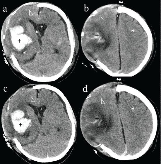
Initial CT findings of left frontoparietal extra-axial hyperdensity and ...
e day after embolization, we performed craniotomy for the left ...
Patient Suffering from Fulminant Guillain-Barré Syndrome Successfully ...
(A) Patient 3: SDH 2 days after excision of the left frontal convexity ...
Management and outcome of spontaneous subaponeurotic fluid collections ...
Anatomy Meninges Ventricles and Neurovasculature Flashcards | Quizlet
CT-of-Ischemic-Stroke
Acute-Ischemic-Stroke-CT
Contrast-vs-Non-Contrast-CT
Ischemic-Stroke-CT-Scan-Images
CT-Head-Ischemic-Stroke
MRI-Image-of-Stroke
Acute-Infarct-CT
Ischemic-Stroke-MRI-DWI
CT-Perfusion-Stroke
Hemorrhagic-vs-Ischemic-Stroke-On-CT
CT-Perfusion-Brain
Normal-Non-Contrast-Head-CT
Imaging-in-Acute-Stroke
Acute-Ischemic-Stroke-Contrast-Stain
Brain-CT-Stroke-Ischemic-Right
Stroke-Aging-MRI


![[PDF] Frontal sinus mucocele with intracranial extension associated ...](https://d3i71xaburhd42.cloudfront.net/627db72ec69b02a6f2840f33af205fbfd37b6e43/1-Figure1-1.png)