Please enter url.
Login
Logout
Please enter url.
Ct Scan Of The Abdomen And Pelvis With And Without Contrast | Hot Sex ...
hotzxgirl.com
source
Comments
CT Scan of the Abdomen and Pelvis: With and Without Contrast ...
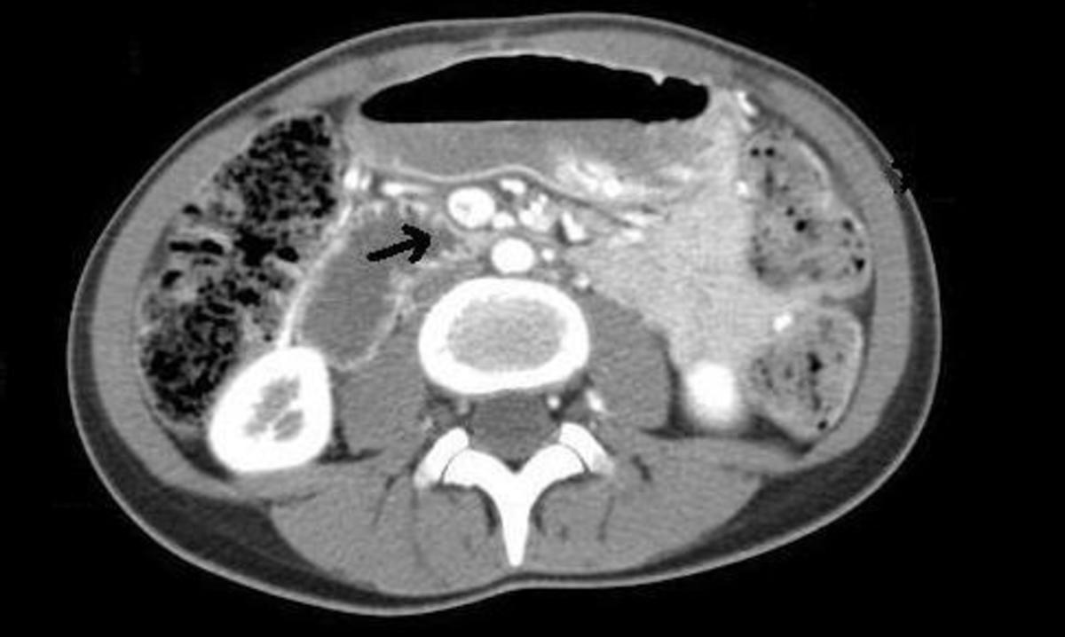
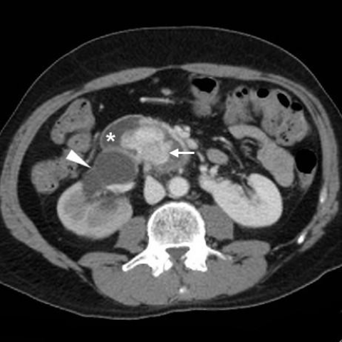
Thickening of the duodenal wall, a pancreatic head compatible with ...
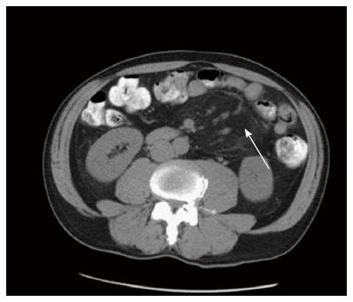
Small bowel faeces sign in patients without small bowel obstruction ...
(PDF) The Anatomic Course of the First Jejunal Branch of the Superior ...
Preoperative CT scan, CTA, and arterial embolization of PRA. (a ...
An abdominal CT scan showed hemoperitoneum from a metastatic ...
Abdominal-pelvic CT-scan. a: Tissular mass of the left pericolic ...
(a) CT of the retroperitoneal mass. Note the vertebral body invasion ...
Lumen Apposing Metal Stent (LAMS) positioned through the posterior ...
Bilateral Supernumerary Kidney: a Very Rare Presentation | Iranian ...
Acute Mesenteric Ischemia: A Challenging Diagnostic Disease—Four Cases ...
Spontaneous bacterial peritonitis complicating decompensated cirrhosis ...
Computed tomography (CT) angiogram with the ruptured splenic artery ...
High-grade small bowel obstruction in an HIV-positive patient | Eurorad
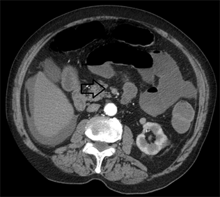
CT Abdomen/Pelvis without contrast. Note the intussusception in the ...
Spontaneous haemoperitoneum from lacerated omental adhesions | Eurorad
Cureus | Macroscopic Hematuria as the Initial Presentation of ...
Superior mesenteric venous thrombosis with small bowel ischemia | Eurorad
Renal carcinoma in horseshoe kidney with endoluminal duodenal invasion ...
Transient, idiopathic adult entero-enteric intussusception | Eurorad
(PDF) Small bowel wall thickening: MDCTevaluation in the emergency room
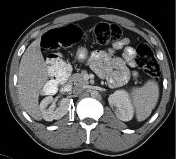
Abdominal CAT scan with oral and iv contrast. The tip of the arrow ...
Internal hernia complicated by small bowel volvulus | Eurorad
Renal carcinoma in a congenital solitary kidney: a therapeutic dilemma ...
Treatment options for spontaneous and postoperative sclerosing mesenteritis
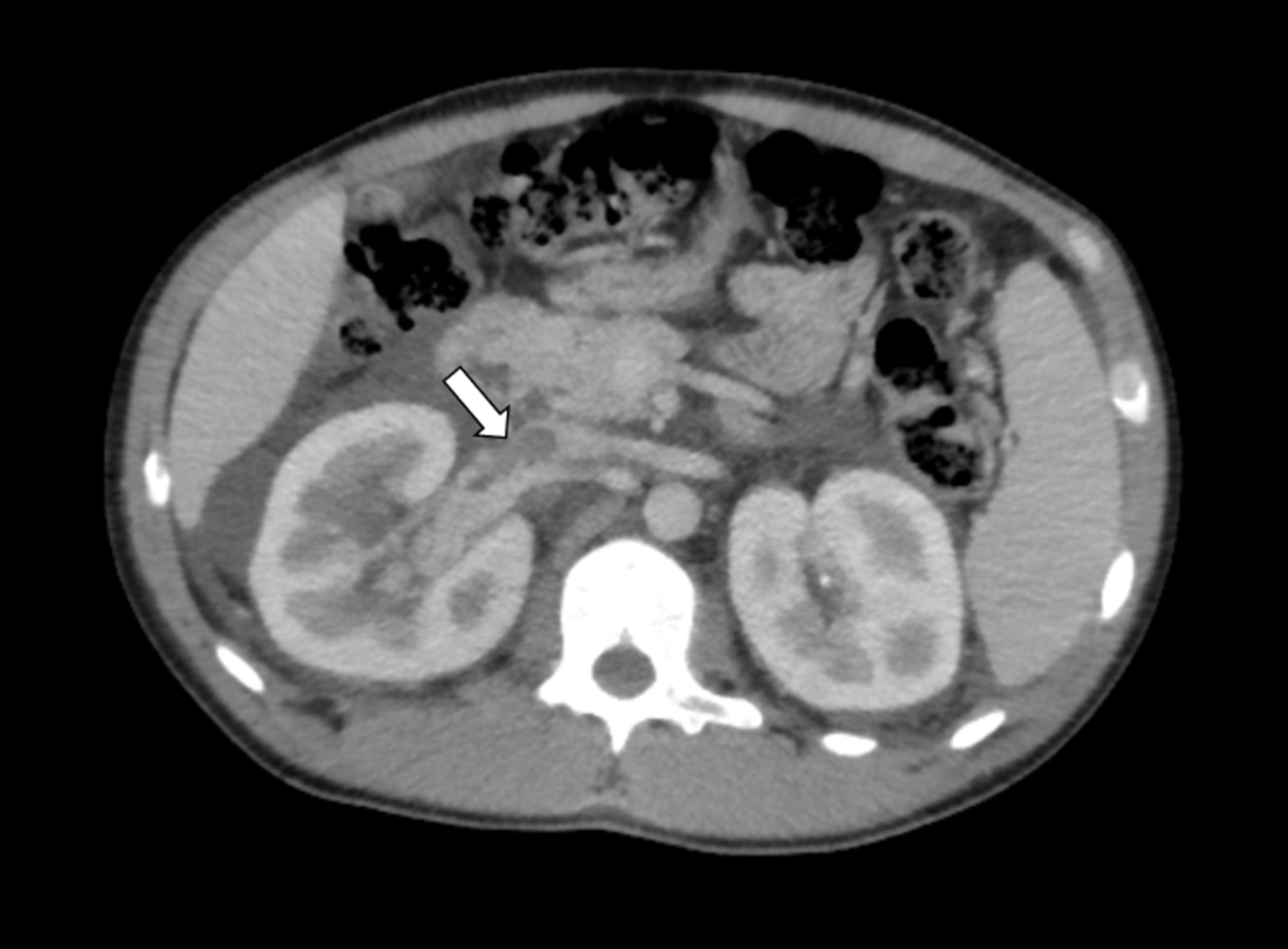
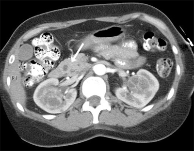
Contrast-enhanced CT shows 6.5×7 cm sized round mass (arrow) with ...
Fig. Signo del anillo hiperdenso en tomografía axial contrastada de ...
Essential thrombocythaemia: abdominal manifestations | Eurorad
Imaging postoperative complications after radical cystectomy with ileal ...
Axial non-contrast CT images through the superior mesenteric vein (A ...
CT of Pheochromocytoma and Paraganglioma: Risk of Adverse Events
A 43-year-old male with Fuhrman grade 2 cystic clear cell renal cell ...
Periportal edema and ascites: computed tomographic signs of traumatic ...
Unusual centrally located abdominal abscess: anatomy as key to the ...
Urology | Basicmedical Key
CT-Abdomen-Pelvis-without-Contrast
Abnormal-CT-Scan-of-Abdomen-and-Pelvis
Abdominal-Hernia-CT-Scan
CT-Abdomen-Pelvis-W-Contrast
Male-Pelvic-CT-Scan
Female-Pelvis-CT-Scan
CT-Scan-for-Abdomen-and-Pelvis
Pelvic-CT-Scan-Cancer
Normal-CT-Scan-Abdomen/Pelvis
CT-Scan-Chest-Abdomen/Pelvis
CT-Abdomen-with-IV-Contrast
Appendicitis-CT-Scan-with-Contrast
CT-Scan-with-Barium-Contrast
MRI-vs-CT-Scan-Abdomen
Abdomen-CT-Scan-Procedure
Abdominal-Aortic-Aneurysm-CT-Scan