Please enter url.
Login
Logout
Please enter url.
HPV- cancer of the base of the tongue. (a) T2w; (b) Post-contrast T1w ...
researchgate.net
source
Comments
Resonancia magnética cerebral de pacientes con diagnóstico de ...
1 EDL tenosynovitis. (A) Longitudinal and (B) transverse US images of ...
a) Sagittal T2 MRI images show cervical spinal epidural hematoma ...
a. T2-weighted MRI of the brain demonstrates an extensive acute ...
A and 3B. Sinonasal cancer in a worker exposed to chromium and nickel ...
Empty sella (left) and tortuosity of the optic nerve (right) in two ...
CT scans of the temporal bone of case 2. A (bone window): a ...
MRI of head and orbits, performed with and without contrast. a Axial ...
Coronal (a) and sagittal (b) MRI with contrast of a patient with a ...
Contiguous sections on T 2-weighted axial MRI illustrating a flow void ...
(A) Initial CT scan indicating dehiscence of the floor of the sella and ...
Pelvic actinomycosis involving the adnexa and uterus in a 68-year-old ...
(a) Axial T 2 weighted MRI reveals a giant schizencephalic cleft and ...
(PDF) Diagnostic Imaging of Peripheral Vestibular Disease in a Chinese ...
Fetal MRI at 27 weeks shows cerebellar hypoplasia (arrow head). An ...
Paranasal magnetic resonance imaging (MRI): (a) left concha bullosa ...
(A) T1-weighted MR image revealed a mass in the right pterygopalatine ...
Craniofacial computed tomography. A. Axial view shows fracture ...
Initial, contrast-enhanced T1-weighted and fat-saturated MRI scan of ...
a,b. Intramedullary intraosseous lipoma in the right nasomaxillary ...
(PDF) Sialoblastoma of the cheek: A case report and review of the ...
(A) Initial CT scan indicating dehiscence of the floor of the sella and ...
Chondrosarcoma. (a) Axial T2-weighted MR image shows a mass with ...
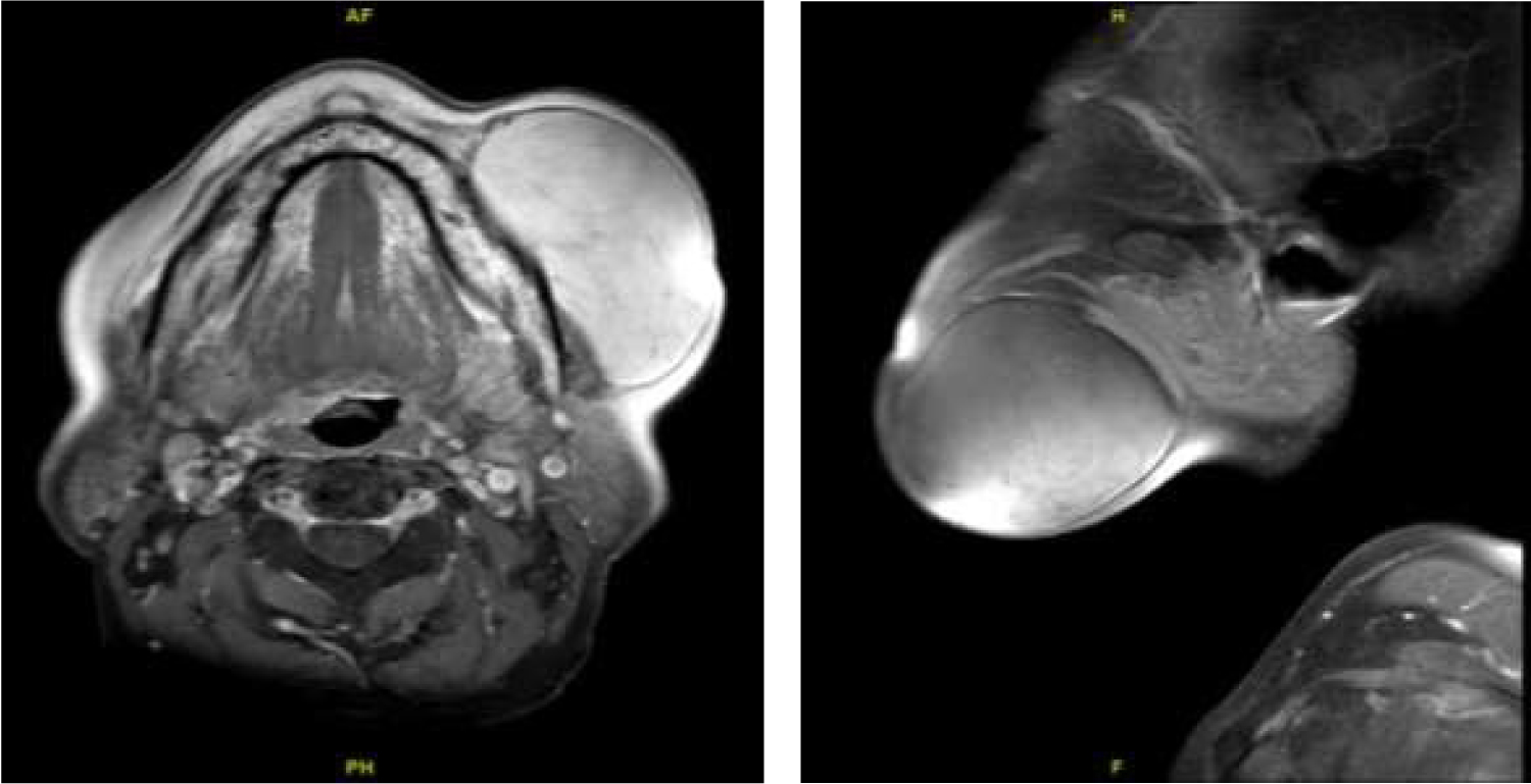
Intra-Oral Approach of Giant Cheek Lipoma Excision Guaranteed Aesthetic ...
Micro CT images after CI in guinea pigs: (A) Sagittal plane of the left ...
Nasal endoscopy of Case 2. Nasal endoscopy showed a bloody tumor mass ...
CT and MRI images of the mandibular region (scale bar, 5 cm). a A CT ...
A) T2 weighted MRI axial view of the brain showing post-surgical tumor ...
Orbital MRI images, axial (a) and coronal (b), revealed enlargement of ...
A Radiologist’s Guide to the 2021 WHO Central Nervous System Tumor ...
Orbital Xanthomas - EyeWiki
Initial magnetic resonance imaging (MRI) demonstrating the hypoplastic ...
Two patients with DIPG who underwent T1-weighted MRI with contrast: (a ...
(PDF) Radiation-induced temporo-mandibular joint disorder in post ...
Patient with acute sensory ataxia related to vitamin B12 deficiency ...