Please enter url.
Login
Logout
Please enter url.
Early experience with feasibility of balloon sinus dilation in ...
onlinelibrary.wiley.com
source
Comments
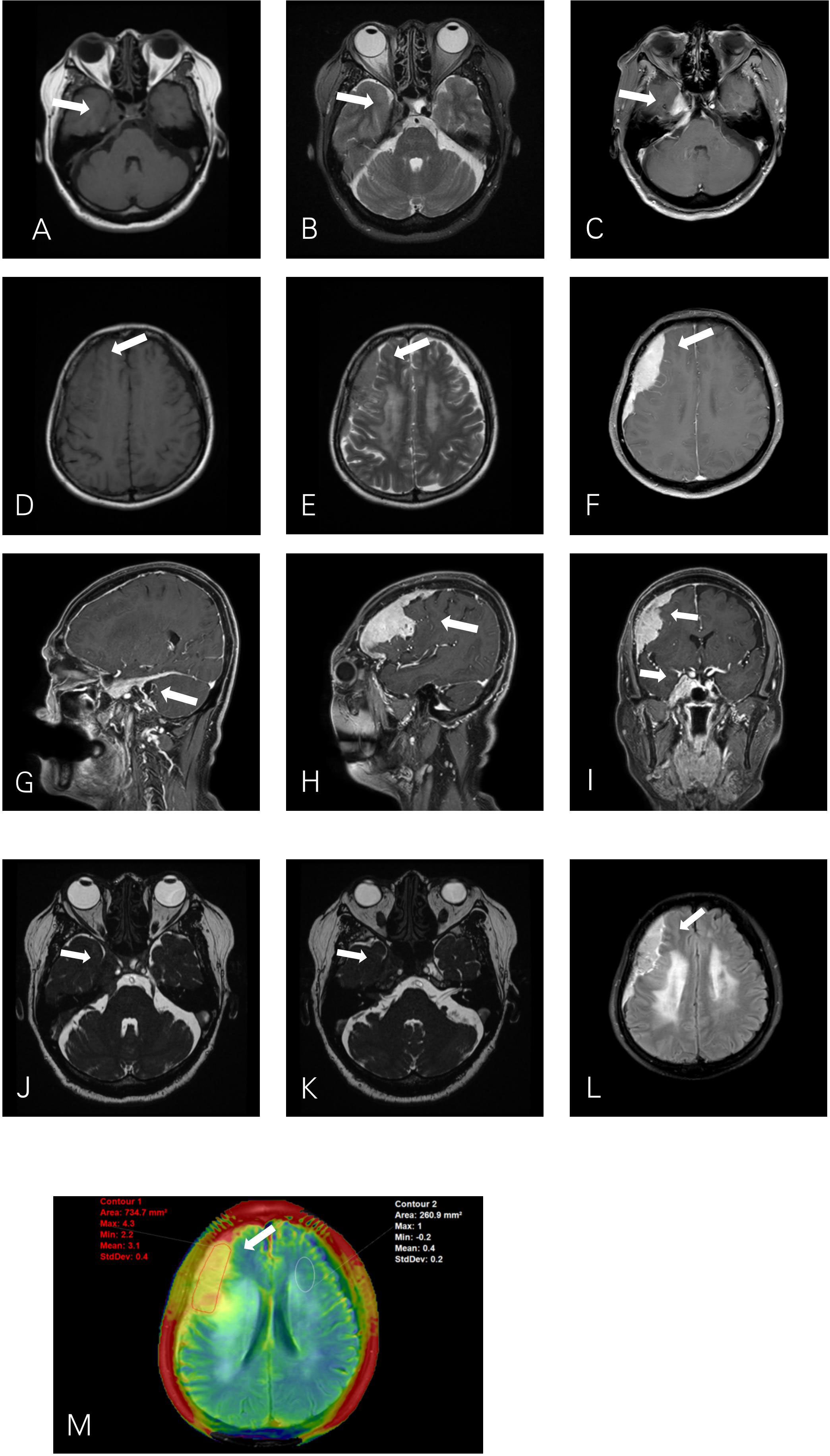
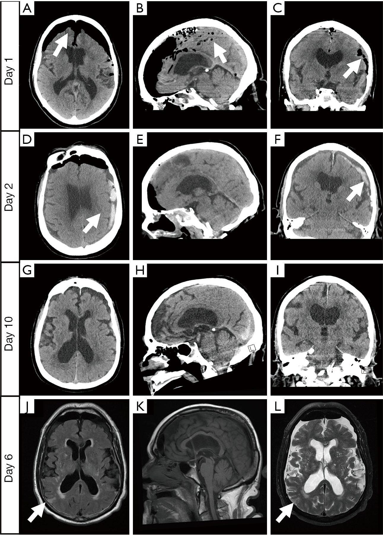
Representative slices from CT and MRI studies. A‐C, Axial, coronal, and ...
Fatal Second Impact Syndrome in Rowan Stringer, A 17-Year-Old Rugby ...
Frontiers | Case report: Primary intracranial mucosa-associated ...
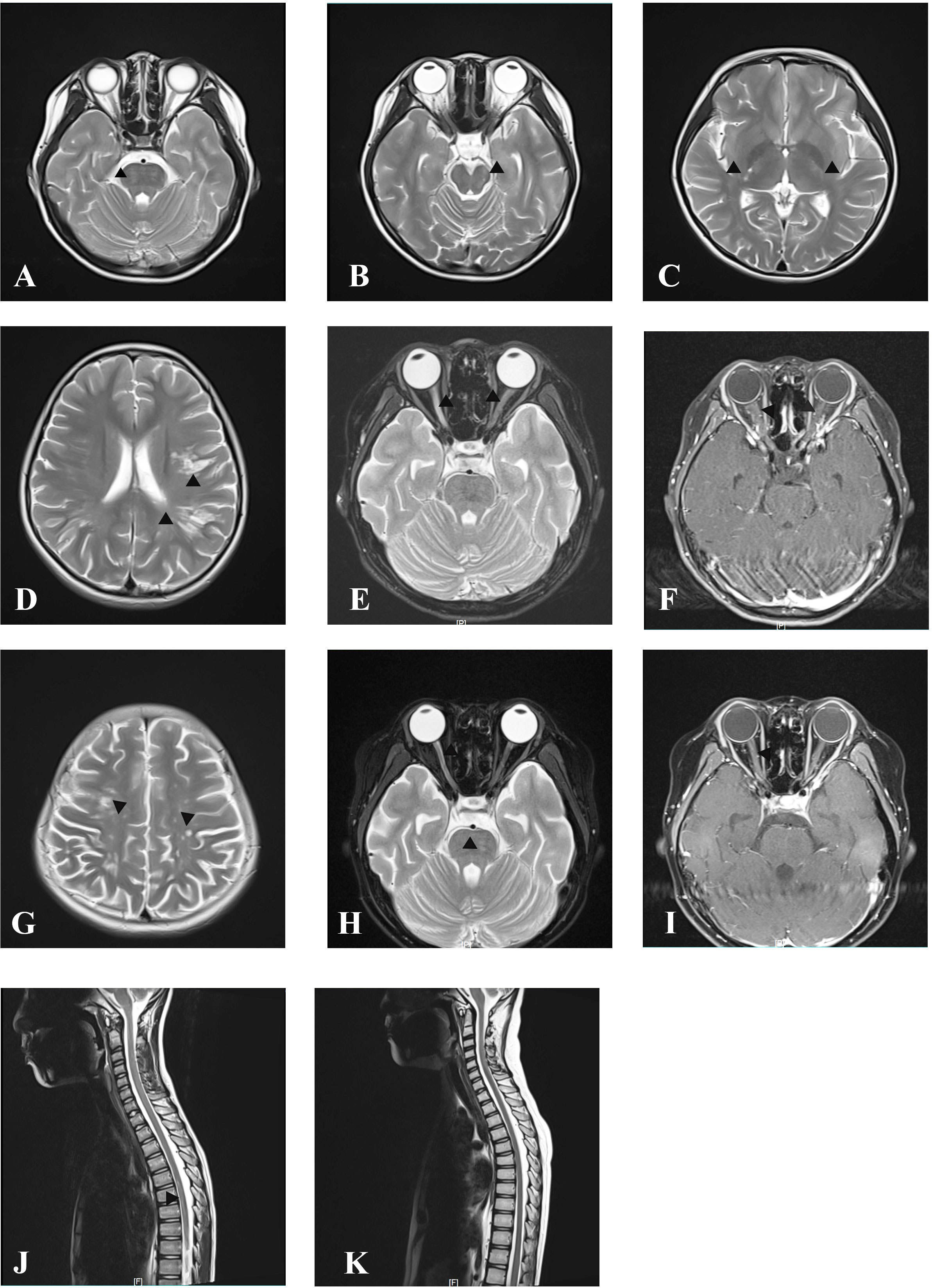
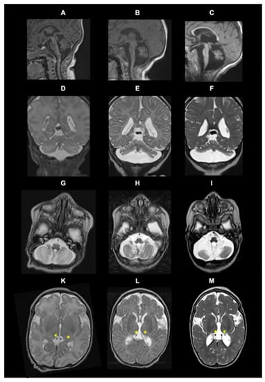
Brain Magnetic resonance imaging of the patient. (A) sagittal ...
Associations of paediatric demyelinating and encephalitic syndromes ...
MACF1 Mutations Encoding Highly Conserved Zinc-Binding Residues of the ...
Figure 3 from Unsupervised Medical Image Translation Using Cycle-MedGAN ...
Figure 2 from Flying focal spot (FFS) in cone-beam CT | Semantic Scholar
Spenden Oberst Blendung west syndrome mri Auto Entfernt Draussen
Brain MRI of patient 1: signal abnormalities in the posterior limbs of ...
Frontiers | Satralizumab as an add-on treatment in refractory pediatric ...
CNTNAP1-Related Congenital Hypomyelinating Neuropathy - Pediatric Neurology
Figure 2 from A Disentangled Representation Based Brain Image Fusion ...
Late-adult onset Leigh syndrome - Journal of Clinical Neuroscience
Figure 1 from A Case of Idiopathic Infratentorial Superficial Siderosis ...
Curves of PSNR values for the 20 MR images. | Download Scientific Diagram
Management of orbital invasion in esthesioneuroblastoma: 14 years ...
Magnetic resonance images of the frontal-perisylvian PNH–PMG subtype ...
Modern Radiation Therapy for Extranodal Nasal-Type NK/T-cell Lymphoma ...
Meningoencephalitis or meningitis in relapsing polychondritis: Four ...
Do Normal D-dimer Levels Reliably Exclude Cerebral Sinus Thrombosis ...
Axial MRI images show areas of abnormal high SI on T2WI (A&B) and FLAIR ...
Frontiers | A Novel CCM2 Gene Mutation Associated With Cerebral ...
Brain-Imaging Features in Individuals with TBCK-Related Encephalopathy ...
Suggestive magnetic resonance imaging (MRI) findings in patients with ...
| CT/MRI findings of patients. (A) MRI demonstrated brainstem malacia ...
Case 2. Immediate postoperative CT scanning consisted of axial (A ...
Figure 1 from Medial posterior choroidal artery territory infarction ...
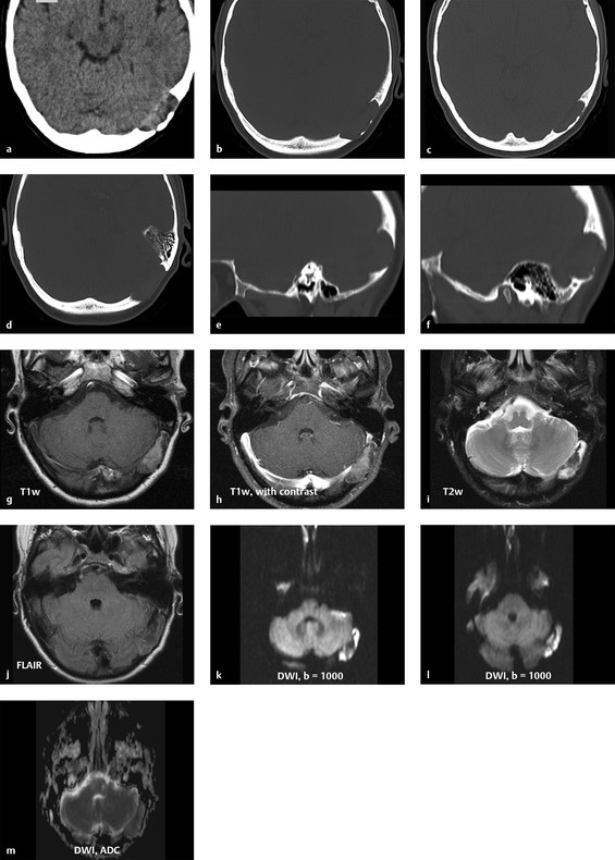
Nothing to sneeze at: tension pneumocephalus causing an acute stroke ...
2.2 Osteolytic Lesions and Lesions with Mixed Features | Radiology Key
Treatment-Related Noncontiguous Radiologic Changes in Children With ...
JCM | Free Full-Text | Pontocerebellar Hypoplasia Type 1D: A Case ...
Sample normal and abnormal brains from the Harvard repository, clinical ...
Figure 1 from MR imaging of epidermoids at the cerebellopontine angle ...
Classic lissencephaly caused by mutations in TUBG1 mutations (A–E ...