Please enter url.
Login
Logout
Please enter url.
Ankylosing Spondylitis Mri Si Joint
mavink.com
source
Comments
Ankylosing Spondylitis | Concise Medical Knowledge
Magnetic resonance imaging (MRI) on diagnosis of juvenile ankylosing ...
Initial (a) and 3.5-year follow-up (b) left parasagittal T2-weighted ...
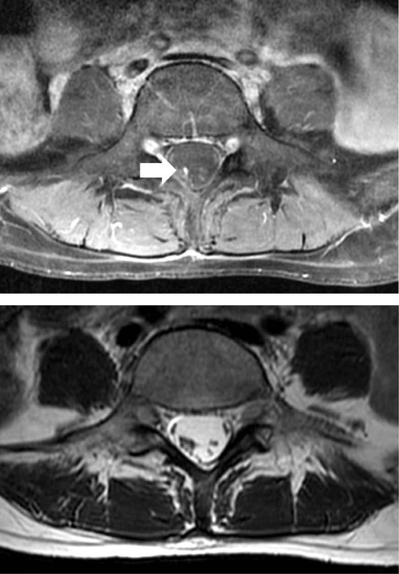
Thoracolumbar spine magnetic resonance imaging (MRI), T1-weighted ...
Coronal oblique STIR image of the sacroiliac joints of a 15-year-old ...
Status post L4–5 and L5–S1 fusion. Spacer cage is displaced ...
Primary Neoplasms | Radiology Key
Facet Arthropathy - Aging Spine - Mitch Medical
RCC intracystic nodules observed using MRI. Case numbers correspond ...
a, b: Vertebral artery loop formation at level of the C6 nerve root on ...
Repeat epidural blood patch at the level of unintentional dural ...
Filum Terminale Mri
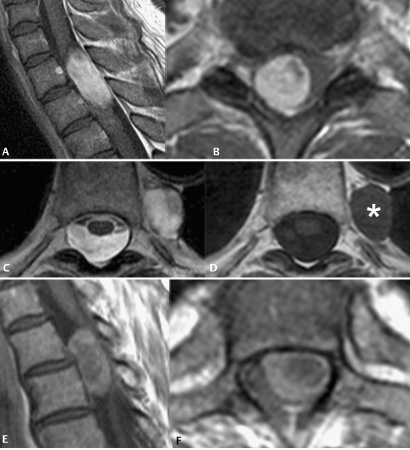
A) Sagittal and (B) axial T1-weighted, and (C) sagittal and (D) axial ...
(a) Posterior endoscopic foraminotomy showing exiting cervical nerve ...
Imaging of sacroiliitis: Current status, limitations and pitfalls ...
The anatomy of physics | Basicmedical Key
MRI of a 48-year-old patient with histologically confirmed sactosalpinx ...
Lumbar spine MRI in upright position for diagnosing acute and chronic ...
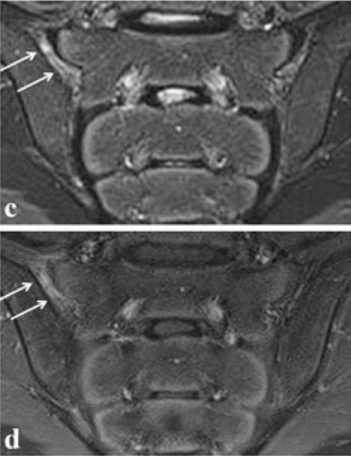
Active sacroiliitis in a 14-year-old (left) and 13-year-old (right) boy ...
Sagittal and axial magnetic resonance imaging in case 1 showing. (A, B ...
Top 3 Spinal Tumors of Each Compartment | Radiology Key
Fast spin echo (FSE) T2-weighted magnetic resonance images (MRI) in the ...
Magnetic Resonance Imaging and Clinical Features in Acute and Subacute ...
Common Spinal Tumors Outside the Top 3 Lists (in Alphabetical Order ...
Lumbosacral conjoined nerve root | Eurorad
Sagittal T2W image (A) of the T11-S1 vertebral column; transverse T2W ...
-Extensive tophaceous gout along the left posterior elements from L3 to ...
a Preoperative sagittal MR image showing a ruptured L4-5 disc with ...
Cervical cord compression with lower limb sensory disturbance: three ...
T2-weighted magnetic resonance imaging (MRI) of Lumbar spine. Sagittal ...
Midsagittal T2-weighted image (A), transverse T2-weighted image at C6-7 ...
Magnetic resonance imaging of dorsal spinal cord. (A) MRI dorsal spine ...
| Images of a female spayed, 6-year-old, mixed breed dog with an acute ...
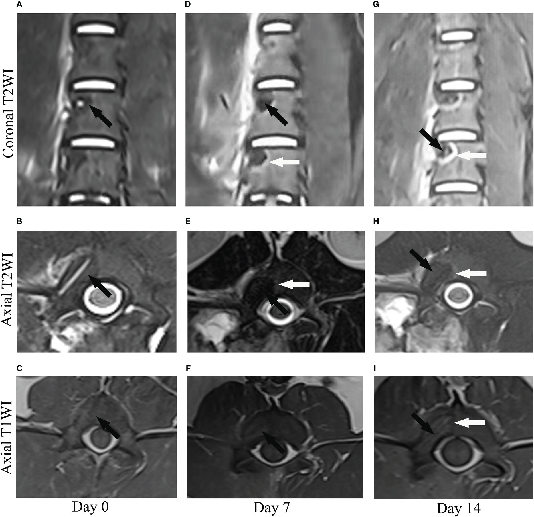
Frontiers | Direct and indirect damage zone of radiofrequency ablation ...
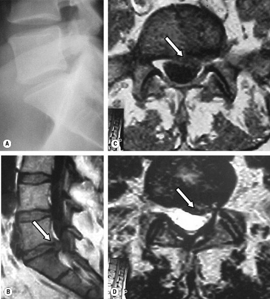
Preoperative T2 sagittal (A, B) and thin section T1 and T2 axial (C, D ...
Ankylosing-Spondylitis-Spine-MRI
Ankylosing-Spondylitis-Lumbar-MRI
Ankylosing-Spondylitis-SI-Joint
Ankylosing-Spondylitis-Kyphosis
Ankylosing-Spondylitis-Hip-Pain
Sacroiliac-Joint-MRI-Scan
Ankylosing-Spondylitis-Neck-MRI
Ankylosing-Spondylitis-CT-Scan
Ankylosing-Spondylitis-X-ray-Findings
Ankylosing-Spondylitis-as-Symptoms
Ankylosing-Spondylitis-MRI-Pelvis
Ankylosing-Spondylitis-X-ray-Images
Sacroiliitis-X-Ray-Findings
Ankylosing-Spondylitis-vs-Spondylosis
Abnormal-SI-Joint-MRI
Andersson-Lesion