Please enter url.
Login
Logout
Please enter url.
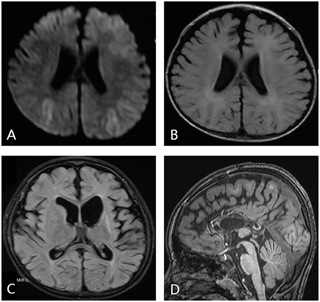
MRI brain. (A) Diffusion weighted image-revealing restriction of ...
researchgate.net
source
Comments
MRI brain. (A) Diffusion weighted image-revealing restriction of ...
Infections of the Developing and Mature Nervous System | Radiology Key
| Brain MRI. Axial T2 (A-C) and diffusion (D-F) showing swelling and ...
Diffuse white matter alterations. Example of typical imaging findings ...
First MR imaging scan (6th month after the initial clinical ...
A typical neuroimage findings: (A) In patient 2, hyperintensities ...
Frontiers | Pediatric epilepsy surgery in patients with Lennox-Gastaut ...
MR images obtained on day 5. AD, On axial T2WI, the lesions appear ...
A) Sagittal T1- weighted magnetic resonance imaging showing spontaneous ...
(PDF) Isolated Sulfite Oxidase Deficiency: Response to Dietary ...
Representative images of infarcts in multiple cerebrovascular ...
Claude's Syndrome Associated with Neurocysticercosis – ScienceOpen
Cerebellitis. A 19-year-old primigravida presented 4 days after labor ...
Cerebral Venous Thrombosis. A 28-year-old female was presented with ...
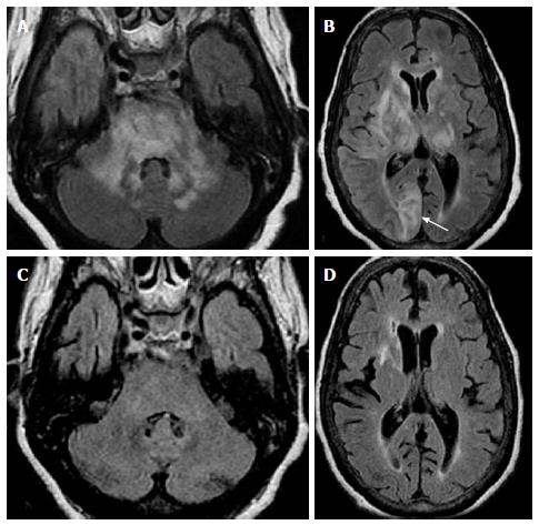
Axial T2 weighted image showing supra- and infra-tentorial high signals ...
Diffusion Weighted Images (DWI); diffuse cortical injury evident by ...
Basal ganglia lesions in children and adults - European Journal of ...
Middle cerebellar peduncles: Magnetic resonance imaging and ...
Cocaine-induced cerebrovascular disease. Magnetic resonance image (MRI ...
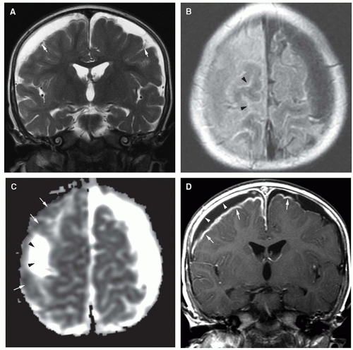
Prechelation MR imaging. A – C , T1- and T2-weighted axial and FLAIR ...
The usefulness of values of the diffusion-weighted imaging (DWI) ratio ...
A , Axial T2-weighted image shows a right frontotem- poral lesion with ...
Restricted Diffusion in Vanishing White Matter | JAMA Neurology | JAMA ...
Manganese accumulation in the brain: MR imaging | SpringerLink
Hemiballism with leg predominance caused by contralateral subthalamic ...
Brain MRI displaying multifocal hyperintense signal changes in ...
The cumulative incidences of all CNS complications and PRES at days 30 ...
Axial (A,B) and coronal (C,D) MRI of the brain/orbits prior to surgical ...
(PDF) A Suspected Case of Cerebral Fat Embolism Triggering a Drug ...
Case 2 at initial presentation. A and B: Axial T2-weighted images show ...
Clinical and genetic analysis of two Chinese infants with Mabry ...
Analysis of Cystic Intracranial Lesions Performed with Fluid-Attenuated ...
Diffusion-Weighted Imaging Abnormalities in Wernicke Encephalopathy ...
MRI (September 20th at 9 p.m.). High signal intensity lesions in ...
MRI images showing the (a) normal appearance of the cerebellum in ...
DWI-MRI-Brain
Diffusion-Tensor
DTI-MRI
Diffusion-Imaging
Tractography-MRI
Facilitated-Diffusion-MRI
DTI-Brain-Scan
Ventriculitis-MRI
Diffusion-Kurtosis-Imaging
Functional-MRI-Brain
DWI-MRI-Sequence
ADC-MRI
Da-Dc-Map-MRI
Acute-Infarct-MRI
Diffusion-MRI-Vision
Apparent-Diffusion-Coefficient