Please enter url.
Login
Logout
Please enter url.
Imaging Inflammation in Acute Brain Ischemia | Stroke
ahajournals.org
source
Comments
Imaging Inflammation in Acute Brain Ischemia | Stroke
Axial magnetic resonance images in a 3-month-old girl to evaluate ...
Patient 1: facial features with down-slanting palpebral fissures ...
Neuroimaging of Cerebrovascular Disease in the Aging Brain
Brain MRI made at the age of 13 months (images A, B, and C) and at the ...
MRI appearance of white matter changes in axial sections of patients ...
Iron Oxide Particle-Enhanced MRI Suggests Variability of Brain ...
Vascular dementia - The Lancet
| Propionic acidemia. Brain MRI: Axial diffusion-weighted imaging (A,B ...
Teaching NeuroImages: Adrenoleukodystrophy presenting as raised ...
Cognitive Outcome at Early School Age in Term-Born Children With ...
MRI of Patient 2, revealing multiple lacunar infarctions of frontal ...
Diffusion-weighted Imaging of Patients with Subacute Cerebral Ischemia ...
Assessment of brain tissue injury after moderate hypothermia in ...
Brain MRI at the second admission. There is an extensive asymmetric ...
Effect of MR sequence on visibility of the pulvinar sign. A ...
Neuroradiological imaging findings. Different severity degrees of brain ...
BIR Publications
Ischemic Stroke From Libman-Sacks Endocarditis Not Associated With ...
Analysis of Cystic Intracranial Lesions Performed with Fluid-Attenuated ...
(a) Non-contrast CT scan showing ill-defined hyperdensi | Open-i
A dominantly inherited mutation in collagen IV A1 (COL4A1) causing ...
Radiographic resolution of an acute silent cerebral ischemic event ...
The Present and the Future of Neuroimaging in Amyotrophic Lateral ...
Cerebral toxoplasmosis in HIV-infected patients over 2015–2018 (a case ...
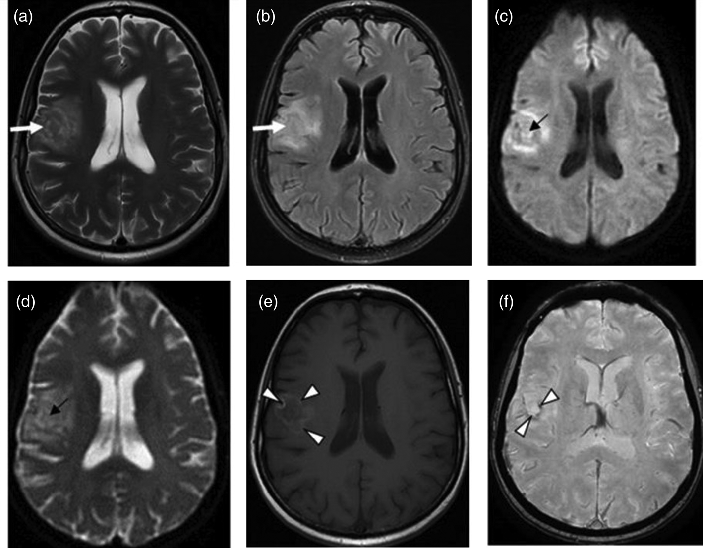
[PDF] Capillary telangiectasias: clinical, radiographic, and ...
Case 27-2018: A 3-Year-Old Boy with Seizures | New England Journal of ...
Reversible encephalopathy caused by an inborn error of cobalamin ...
Brainstem signs with progressing atrophy of medulla oblongata and upper ...
Parasitic Diseases - Diffusion weighted and diffusion tensor imaging: a ...
a Axial T2-weighted and b axial T1-weighted post-contrast images show a ...
Cerebral toxoplasmosis responding to therapy. (A) Axial T2-weighted ...
PMG. Axial T2-weighted image shows microgyria with normal cortical ...
(A, B) Role of structural MRI as an exclusionary tool. Both cases ...
Cerebral MRI, 2 months after onset of psychosis, epilepsy, and visual ...
MRI-Brain-Infarct
Embolic-Stroke-MRI
Diffusion-MRI-Stroke
Abnormal-Brain-MRI
Acute-Stroke-On-MRI
MRI-Brain-Scan
MCA-Stroke-MRI
Brain-Stem-Stroke-MRI
MRI-DWI-Stroke
Hemorrhagic-Stroke-MRI
MRI-Scan-Brain-Stroke-Images
Cerebellar-Stroke-MRI
Subacute-Stroke-MRI
Reading-Brain-MRI
CT-Brain-Stroke
Dementia-Brain-MRI


![[PDF] Capillary telangiectasias: clinical, radiographic, and ...](https://d3i71xaburhd42.cloudfront.net/317f2c6908783a035e3db0f26d7f7100f30ee937/4-Figure3-1.png)


