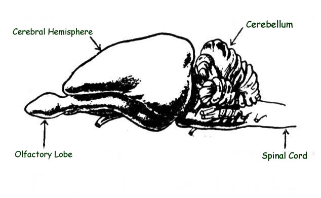
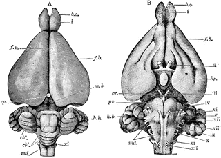
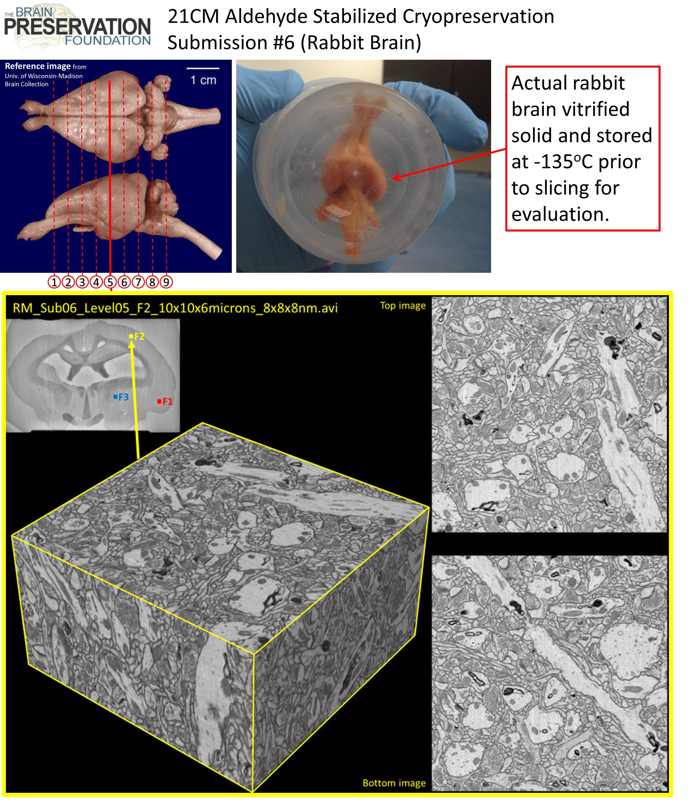
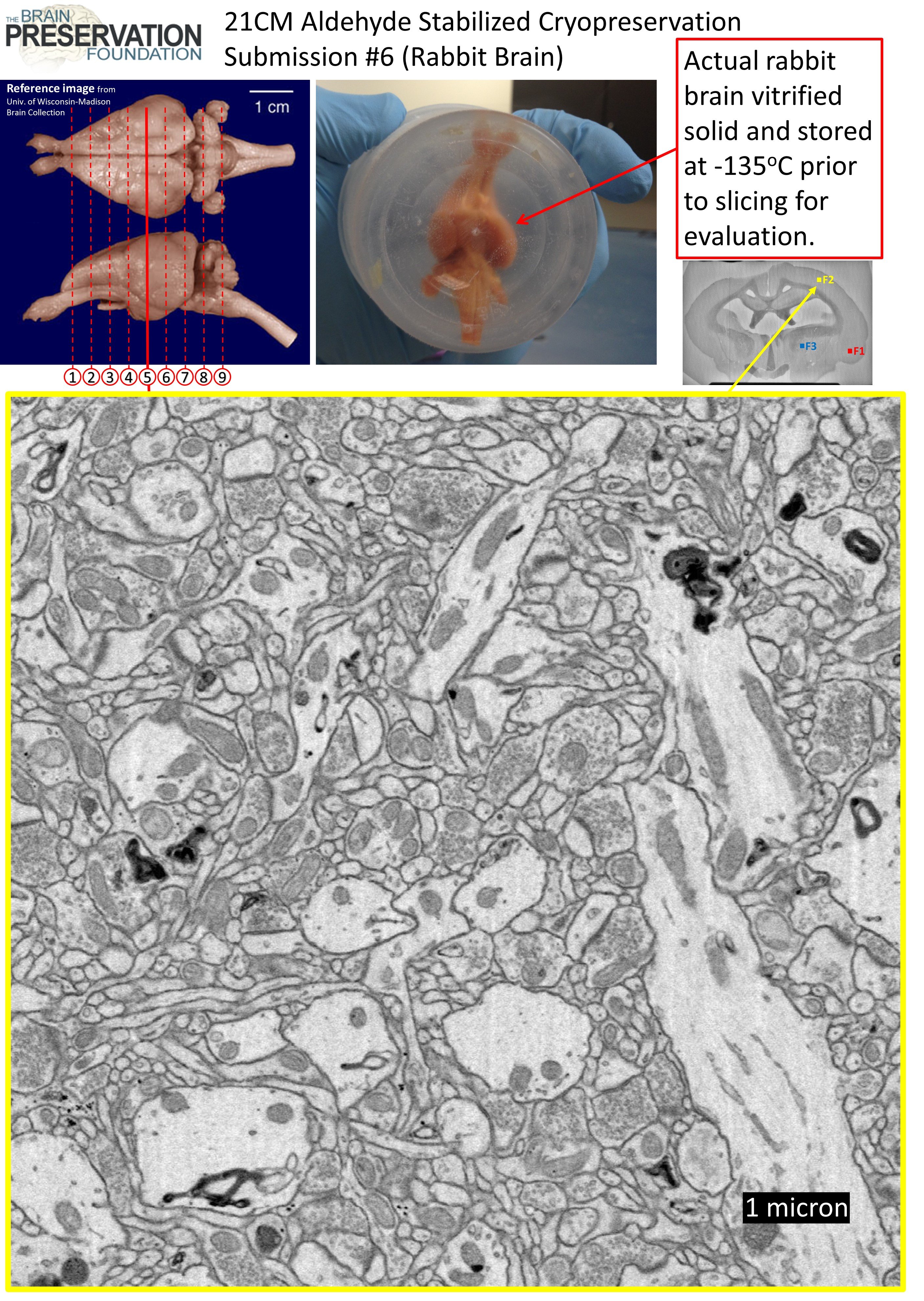
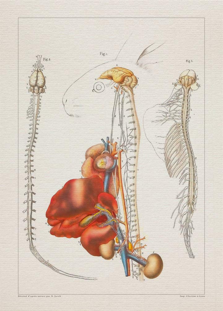
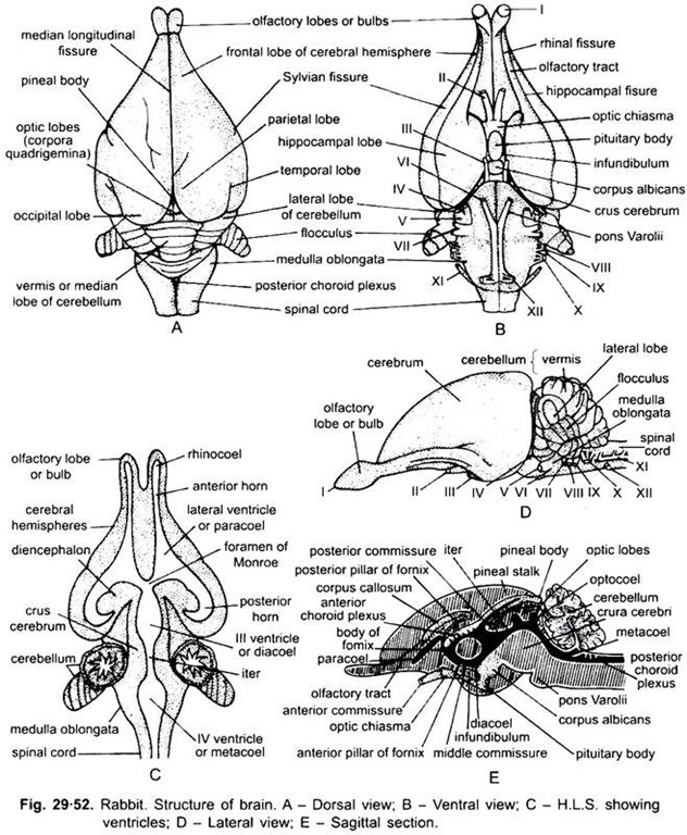
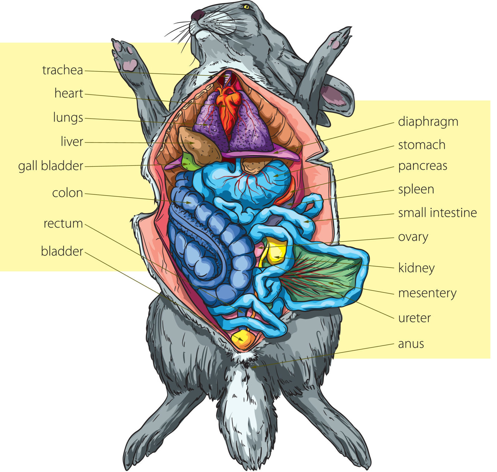
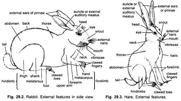
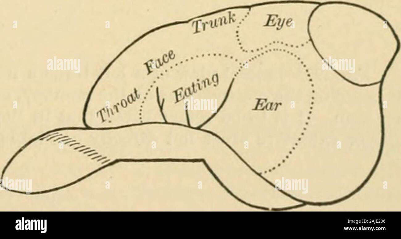
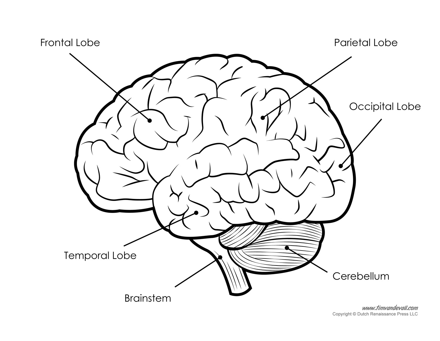
Well Labelled Rabbit Brain


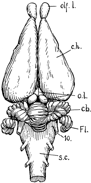


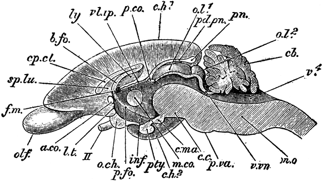


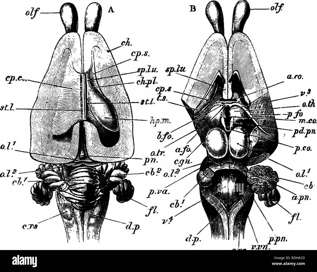


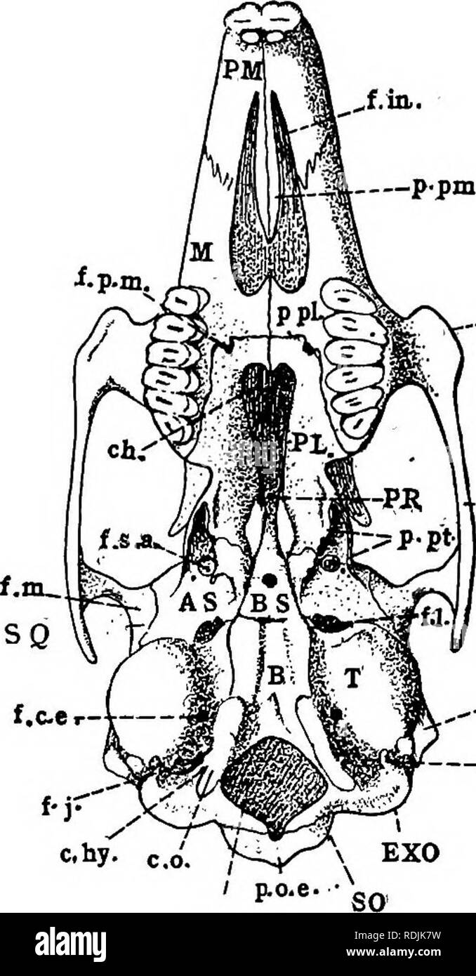


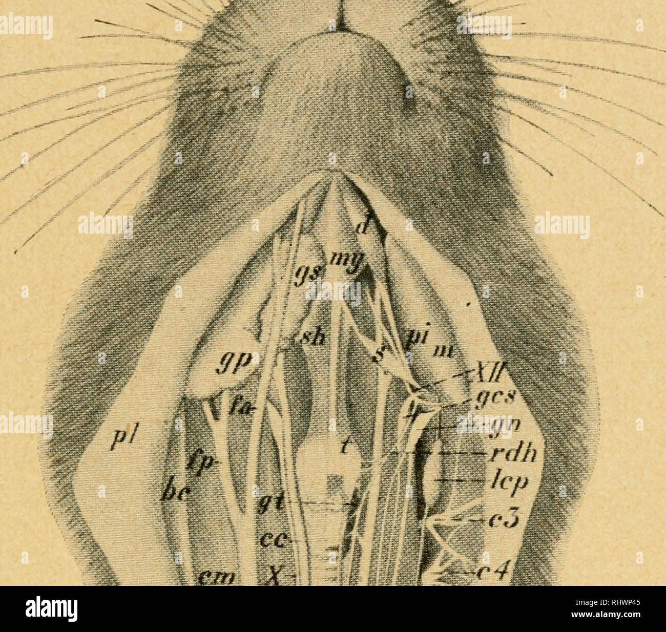


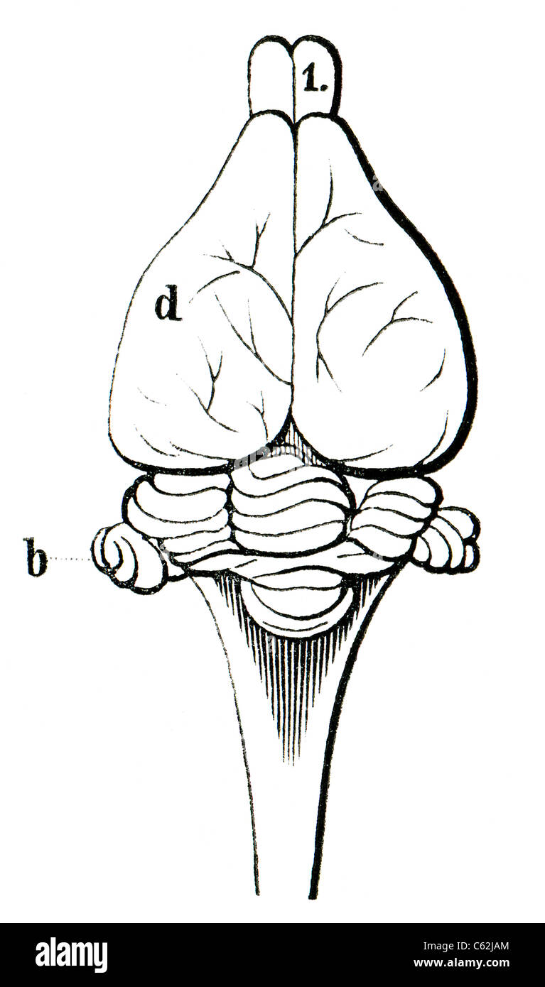


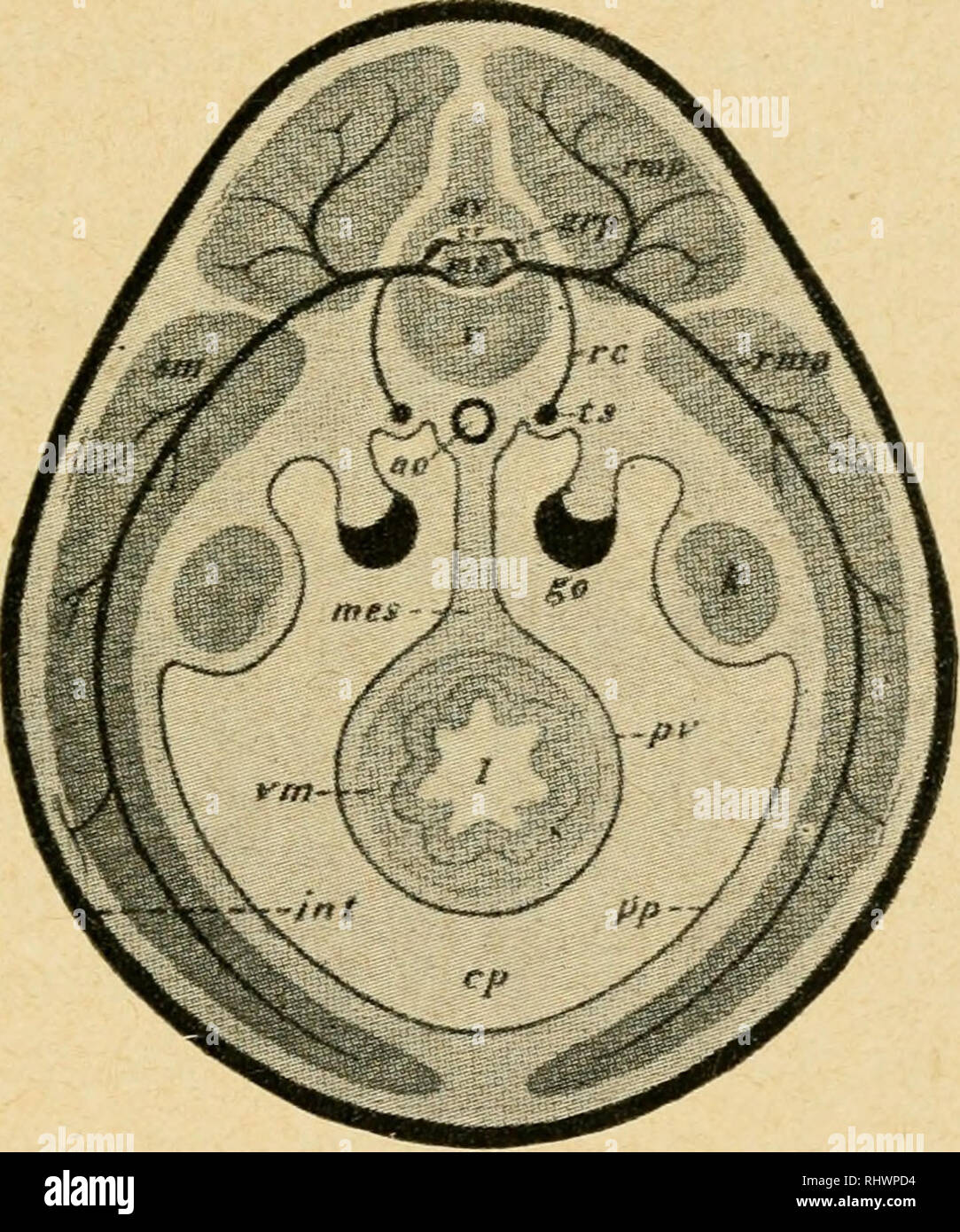
![. Practical anatomy of the rabbit [microform] : an elementary ...](https://c8.alamy.com/comp/RENR0G/practical-anatomy-of-the-rabbit-microform-an-elementary-laboratory-textbook-in-mammalian-anatomy-lapins-anatomy-comparative-rabbits-rabbits-lapins-anatomie-compare-192-anatomy-of-the-rabbit-e-the-posterior-funiculus-in-passing-forward-from-the-cord-is-divided-into-medial-and-lateral-portions-the-medial-portion-the-fasciculus-gracilis-forms-a-narrow-band-ter-minating-forwards-in-a-club-shaped-expansion-the-clava-the-lateral-portion-the-fasciculus-cuneatus-passes-into-the-restiform-body-6-the-brain-may-be-divided-by-a-median-vertical-section-and-one-half-examined-RENR0G.jpg)


Drive innovation through comprehensive galleries of industry-focused Well Labelled Rabbit Brain photographs. showcasing industrial applications of computer, digital, and electronic. perfect for industrial documentation and training. Browse our premium Well Labelled Rabbit Brain gallery featuring professionally curated photographs. Suitable for various applications including web design, social media, personal projects, and digital content creation All Well Labelled Rabbit Brain images are available in high resolution with professional-grade quality, optimized for both digital and print applications, and include comprehensive metadata for easy organization and usage. Our Well Labelled Rabbit Brain gallery offers diverse visual resources to bring your ideas to life. The Well Labelled Rabbit Brain archive serves professionals, educators, and creatives across diverse industries. Reliable customer support ensures smooth experience throughout the Well Labelled Rabbit Brain selection process. Each image in our Well Labelled Rabbit Brain gallery undergoes rigorous quality assessment before inclusion. Time-saving browsing features help users locate ideal Well Labelled Rabbit Brain images quickly. The Well Labelled Rabbit Brain collection represents years of careful curation and professional standards. Cost-effective licensing makes professional Well Labelled Rabbit Brain photography accessible to all budgets. Professional licensing options accommodate both commercial and educational usage requirements. Whether for commercial projects or personal use, our Well Labelled Rabbit Brain collection delivers consistent excellence.