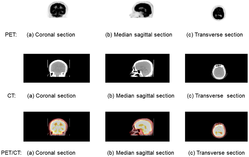
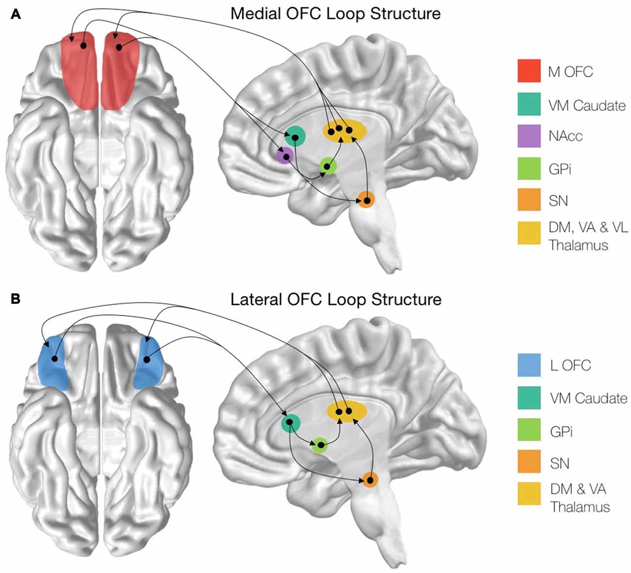
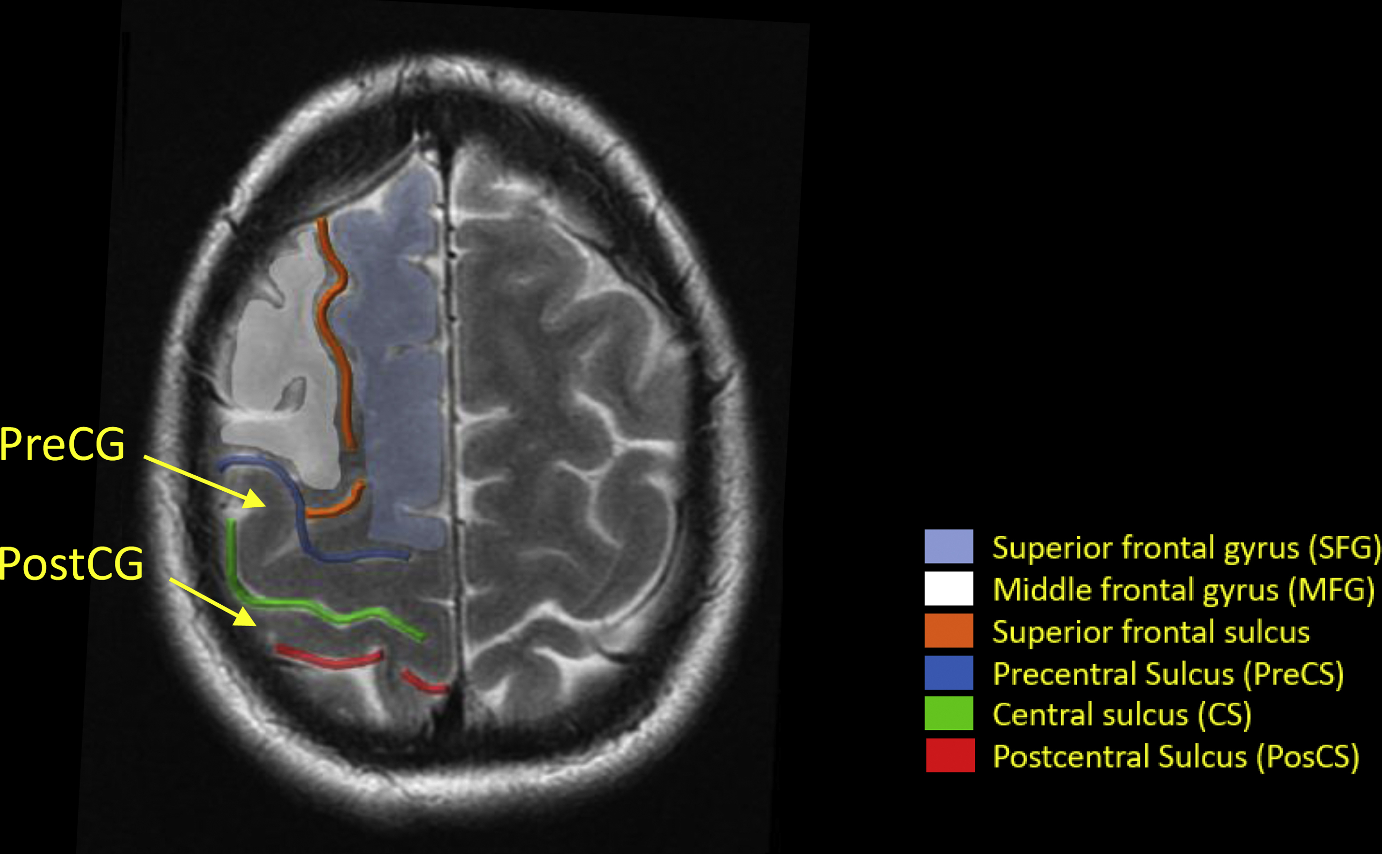
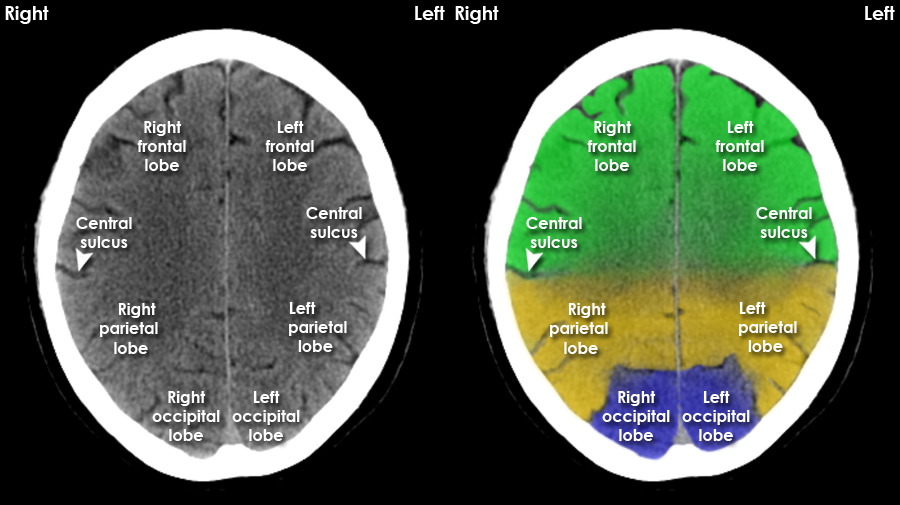
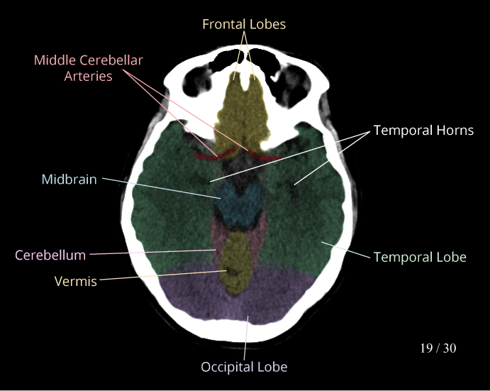
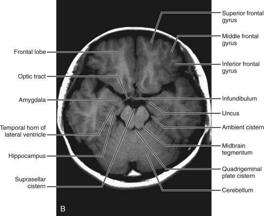
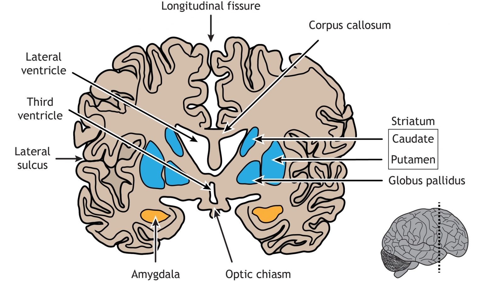
Superior Rontal Cortex Pet/ct


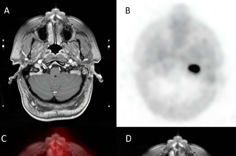


![PET/CT with [ 11 C]MET-frontal projection. Visible focus of increased ...](https://www.researchgate.net/publication/366832577/figure/fig2/AS:11431281111365796@1672943158996/a-PET-CT-with-11-C-MET-frontal-projection-Visible-focal-increased-radiotracer_Q320.jpg)


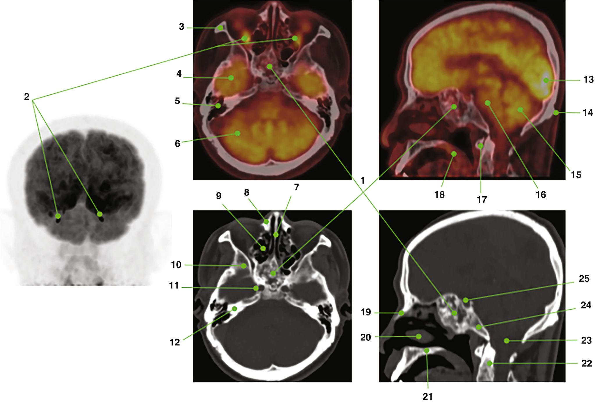
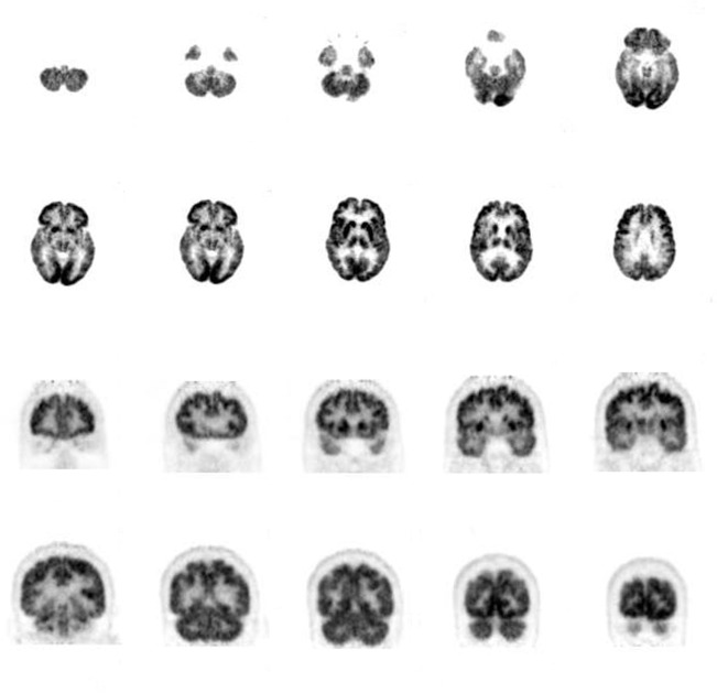
![Brain [¹⁸F]FDG‐PET. Top row shows fused PET and CT axial sections at ...](https://www.researchgate.net/publication/346450019/figure/fig1/AS:1010287088005125@1617882620812/Brain-FFDG-PET-Top-row-shows-fused-PET-and-CT-axial-sections-at-the-level-of-mesial.png)
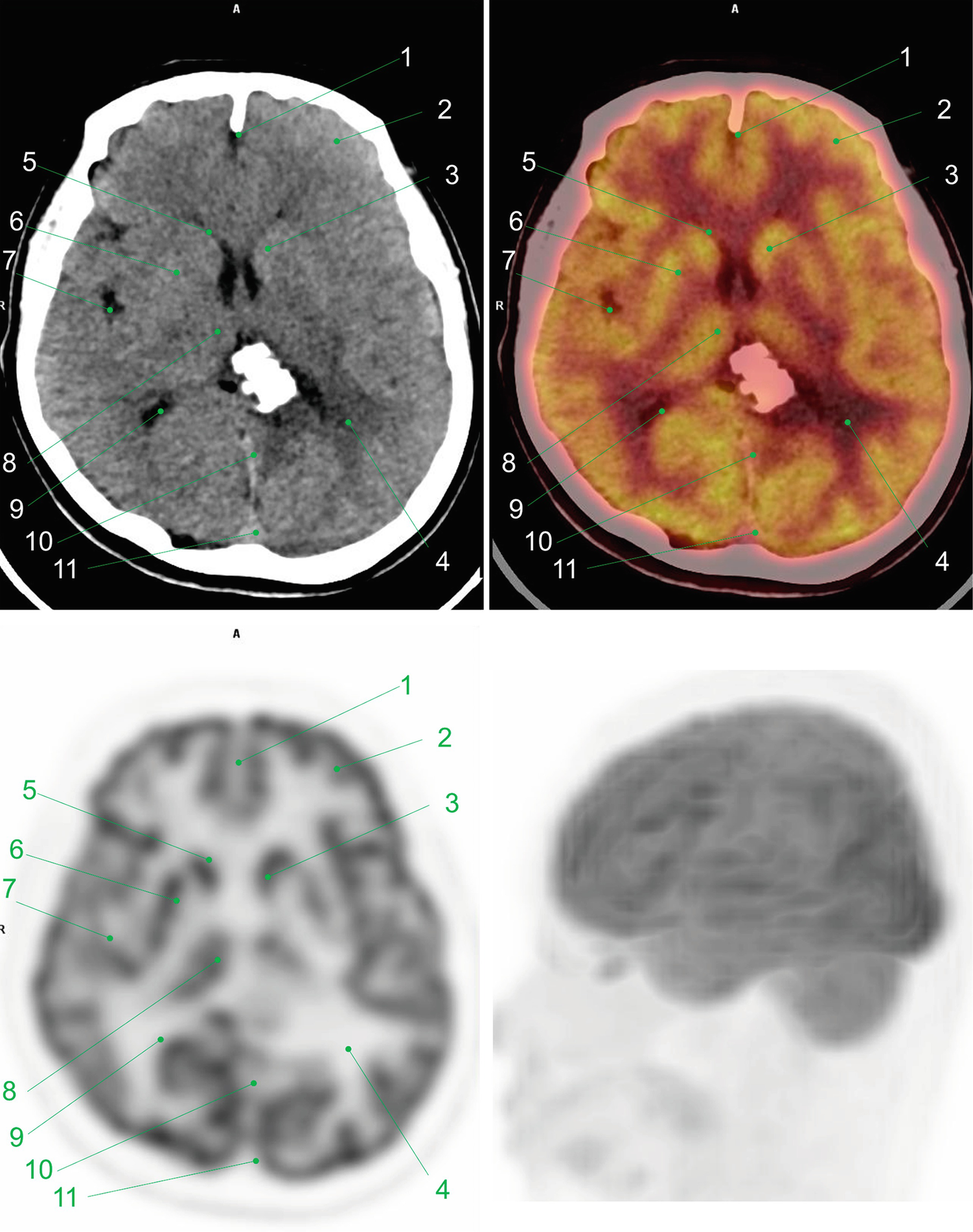
![Figure. Brain [18F]FDG-PET images showing bilateral temporoparietal ...](https://www.researchgate.net/publication/336751681/figure/fig1/AS:820586985029633@1572654591843/Figure-Brain-18FFDG-PET-images-showing-bilateral-temporoparietal-hypometabolism-with.png)


![[번역]인간의 뇌(Human Brain) : 네이버 블로그](https://upload.wikimedia.org/wikipedia/commons/3/35/Lateral_surface_of_cerebral_cortex_-_gyri.png)


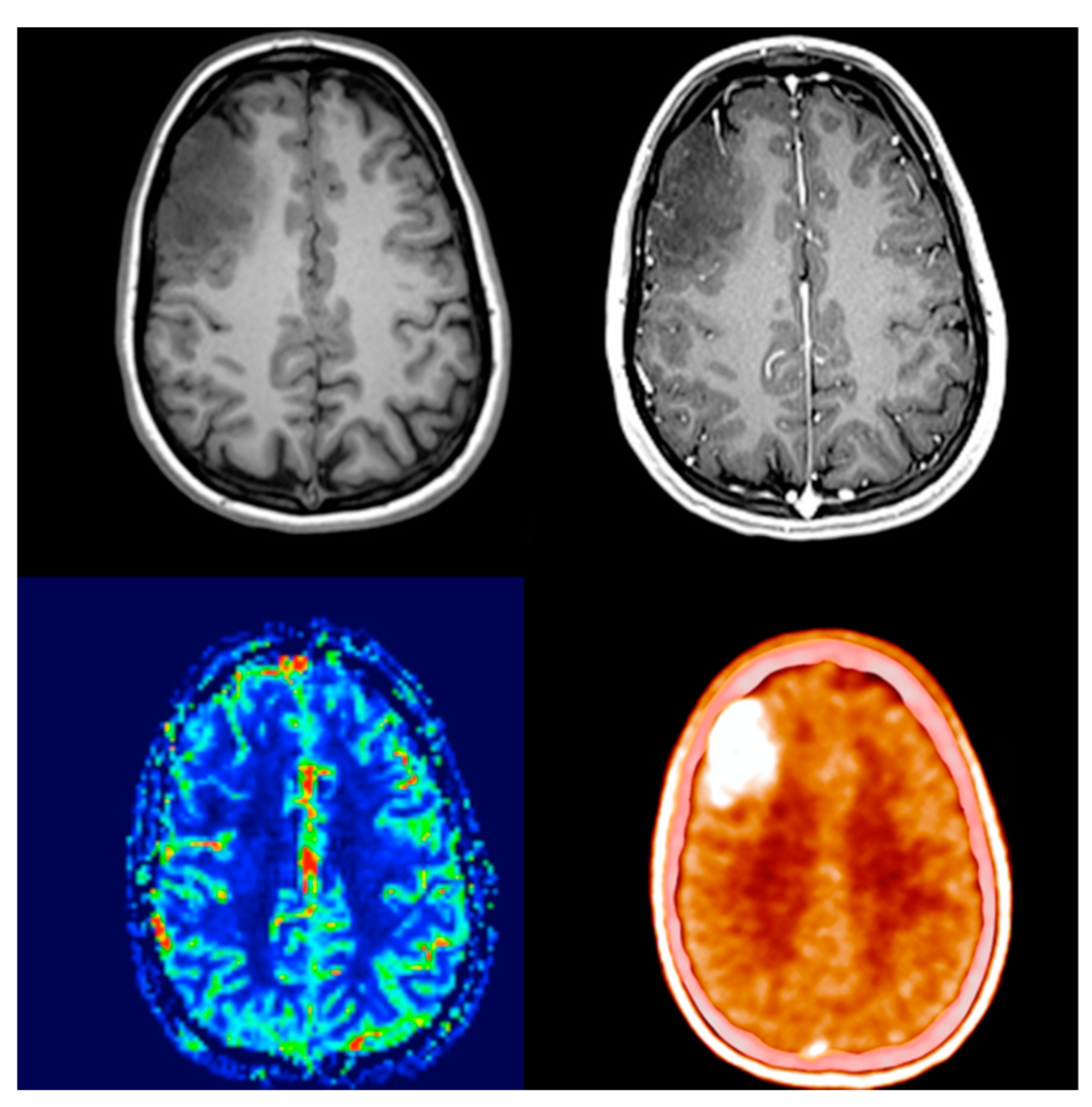
![Caso Clínico PET/RM cerebral 2-[18F] FDG – Grupo CT Scanner](https://grupoctscanner.com/wp-content/uploads/2022/12/deterioro_cognitivo.png)


Discover traditions with our cultural Superior Rontal Cortex Pet/ct gallery of extensive collections of diverse images. preserving heritage via photography, images, and pictures. designed to promote cultural understanding. Discover high-resolution Superior Rontal Cortex Pet/ct images optimized for various applications. Suitable for various applications including web design, social media, personal projects, and digital content creation All Superior Rontal Cortex Pet/ct images are available in high resolution with professional-grade quality, optimized for both digital and print applications, and include comprehensive metadata for easy organization and usage. Our Superior Rontal Cortex Pet/ct gallery offers diverse visual resources to bring your ideas to life. Comprehensive tagging systems facilitate quick discovery of relevant Superior Rontal Cortex Pet/ct content. Whether for commercial projects or personal use, our Superior Rontal Cortex Pet/ct collection delivers consistent excellence. Time-saving browsing features help users locate ideal Superior Rontal Cortex Pet/ct images quickly. Regular updates keep the Superior Rontal Cortex Pet/ct collection current with contemporary trends and styles. Diverse style options within the Superior Rontal Cortex Pet/ct collection suit various aesthetic preferences. The Superior Rontal Cortex Pet/ct archive serves professionals, educators, and creatives across diverse industries. Each image in our Superior Rontal Cortex Pet/ct gallery undergoes rigorous quality assessment before inclusion.