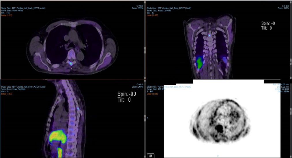
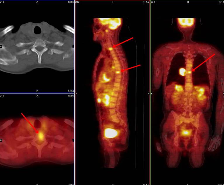
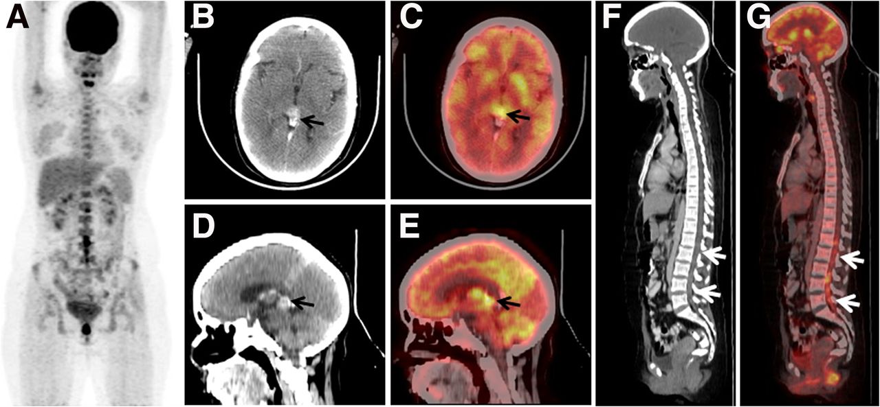
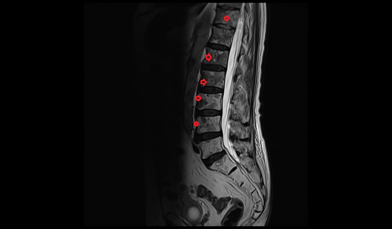
Spinal Metastasis Pet/ct
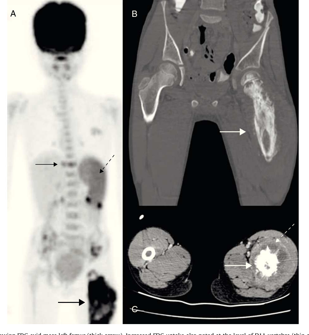


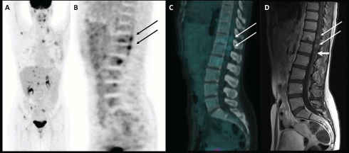


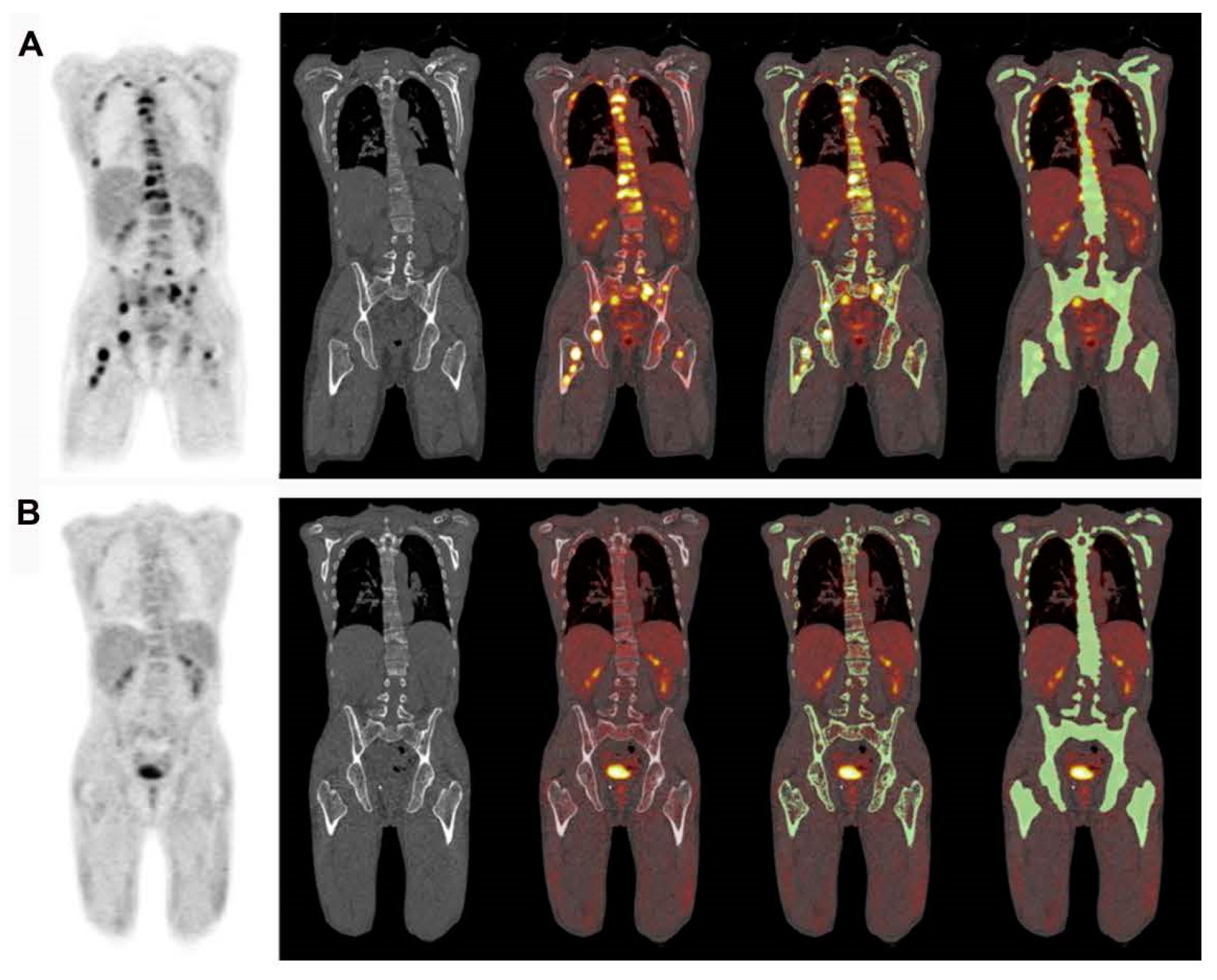
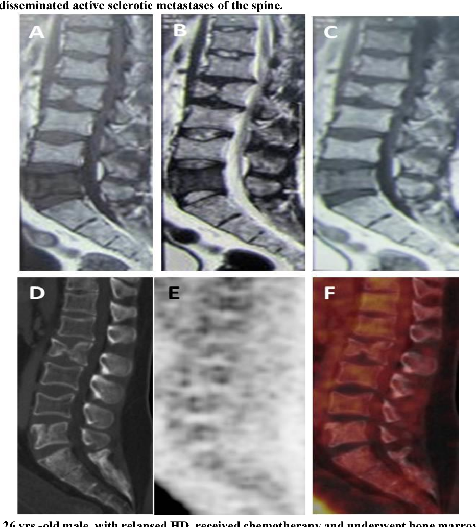
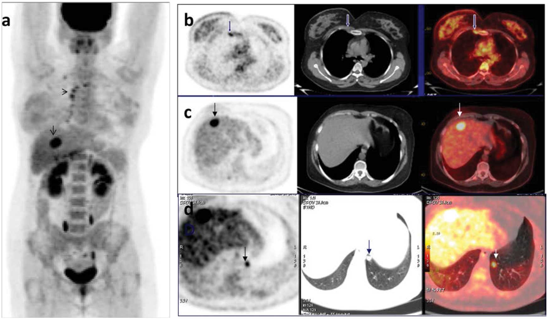
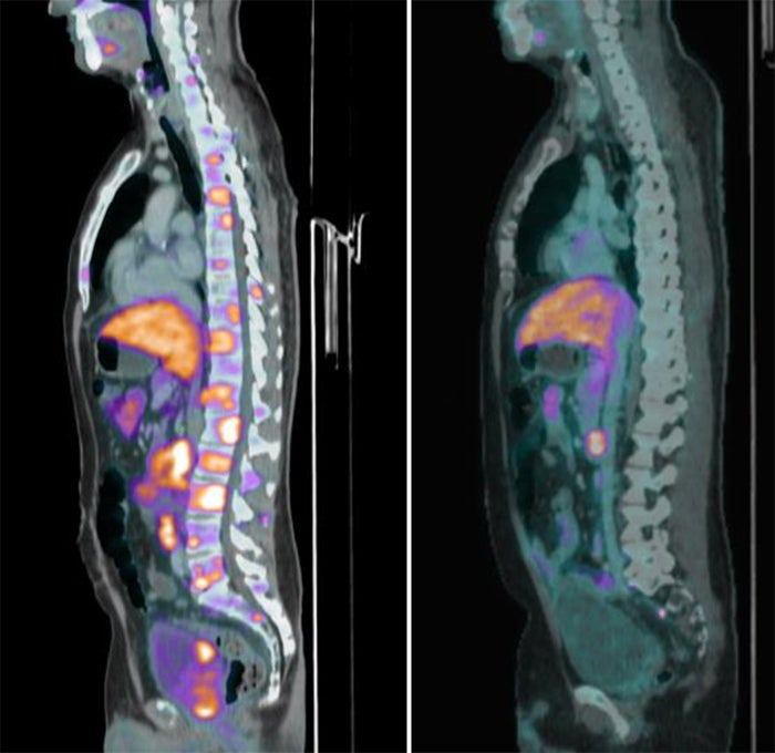
![FDG PET/CT [(A) 7/21] with multiple FDG avid bone metastases (ribs ...](https://www.researchgate.net/publication/366193597/figure/fig1/AS:11431281106831940@1670848655364/FDG-PET-CT-A-7-21-with-multiple-FDG-avid-bone-metastases-ribs-spine-femurs_Q640.jpg)


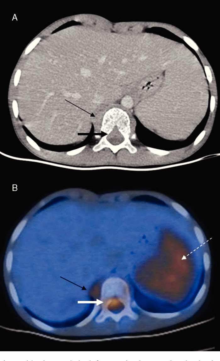
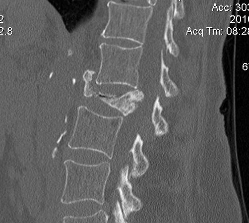
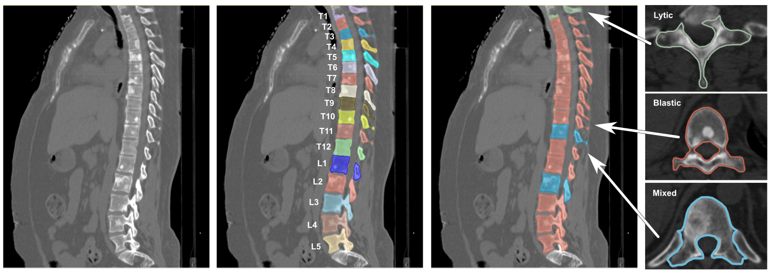
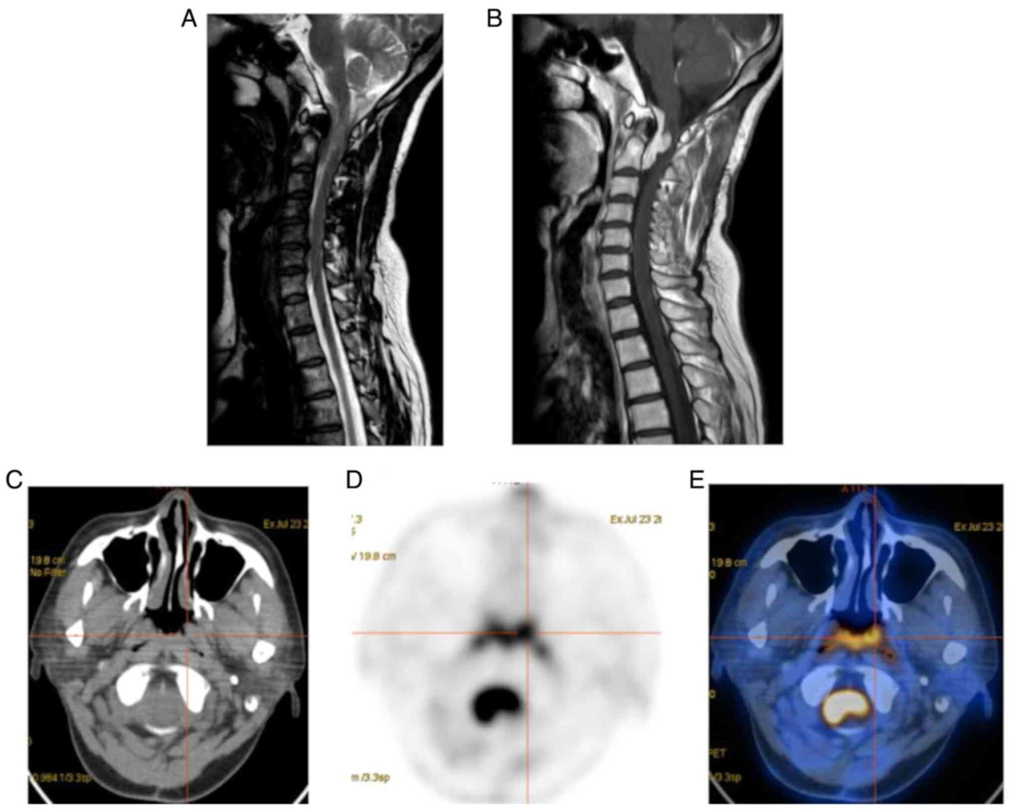
![[Translated article] Imaging diagnosis of vertebral metastasis ...](https://static.elsevier.es/multimedia/18884415/0000006700000006/v3_202403090752/S1888441523001777/v3_202403090752/en/main.assets/gr3.jpeg?xkr=ue/ImdikoIMrsJoerZ+w997EogCnBdOOD93cPFbanNdV4B9vwZQjgKcZv3MP4Cds3qiLBpJmV7qW2dfcCTNKQL+fntRnfj43PfmUd5K4KZbjquQl1mLMSE3KGOyQBDl91pzGZDpOj8la9yAE0kw7viXBSCeobkmYGcuS3L0BzOhQcpbNpVAJA3ppd+IhI2MMtWwUYHLQI1l+RHSuO5gwnV/XmMhodN5Pj9Ioq5aF2AcWZ+khCZQwTJfmqzuhdvy8/gNeRR9t0dLb7kP6bqRP4h0SAe3L89P79w7vOr4Hd0+trogUtHGd0FnNVECeoH1t)


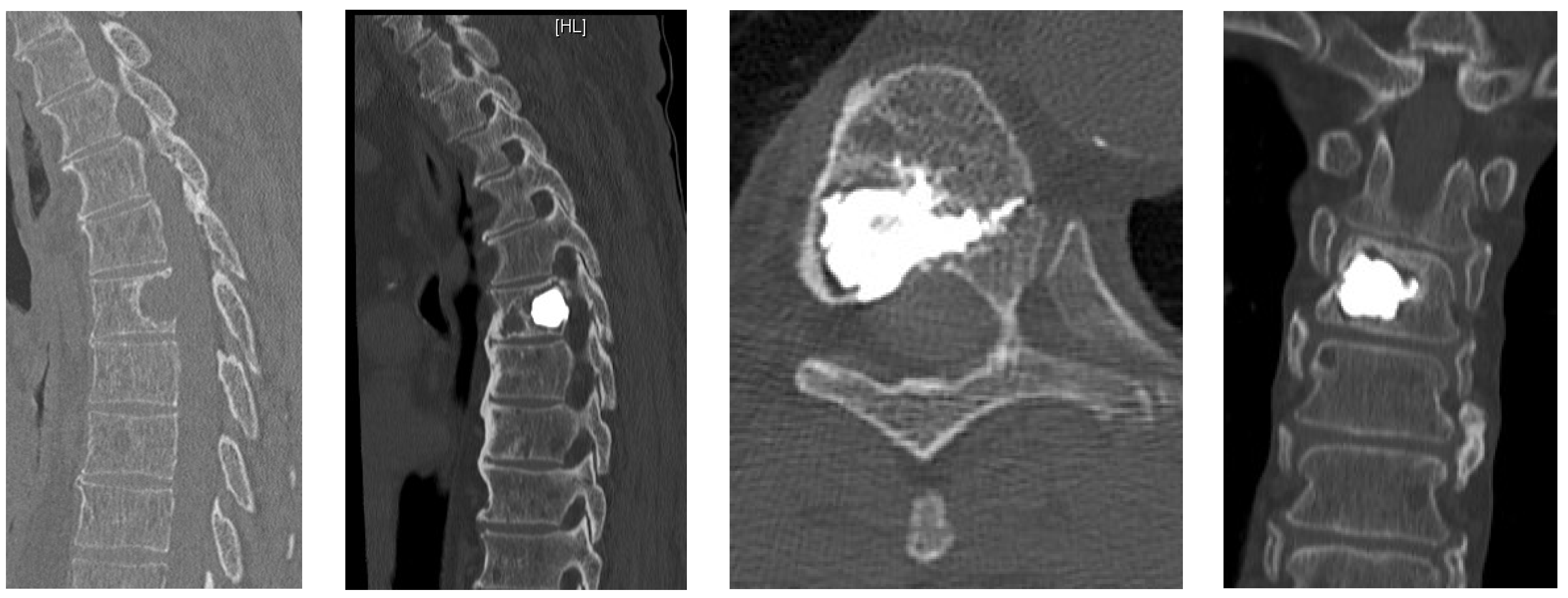
![(PDF) Diagnostic performance of [68Ga]DOTATATE PET/CT, [18F]FDG PET/CT ...](https://i1.rgstatic.net/publication/379867861_Diagnostic_performance_of_68GaDOTATATE_PETCT_18FFDG_PETCT_MRI_of_the_spine_and_whole-body_diagnostic_CT_and_MRI_in_the_detection_of_spinal_bone_metastases_associated_with_pheochromocytoma_and_paragang/links/661f1d5f39e7641c0bd24053/largepreview.png)


![[¹⁸F]-PSMA-1007-PET/CT on a digital scanner illustrates bone metastasis ...](https://www.researchgate.net/publication/352367169/figure/fig7/AS:1034464197083140@1623646892896/F-PSMA-1007-PET-CT-on-a-digital-scanner-illustrates-bone-metastasis-and-UBU-in-two.png)


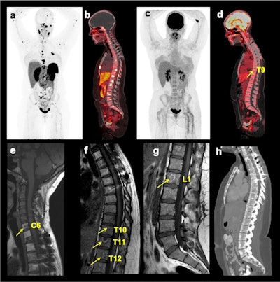
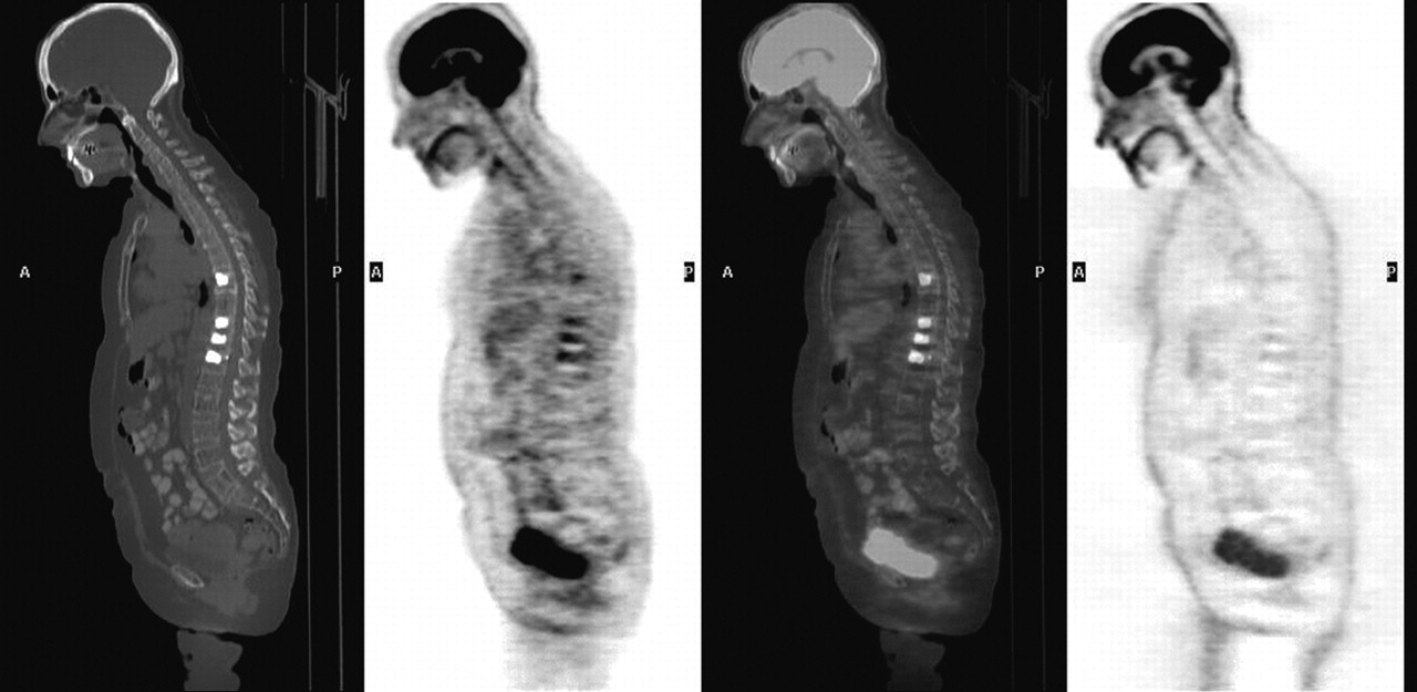
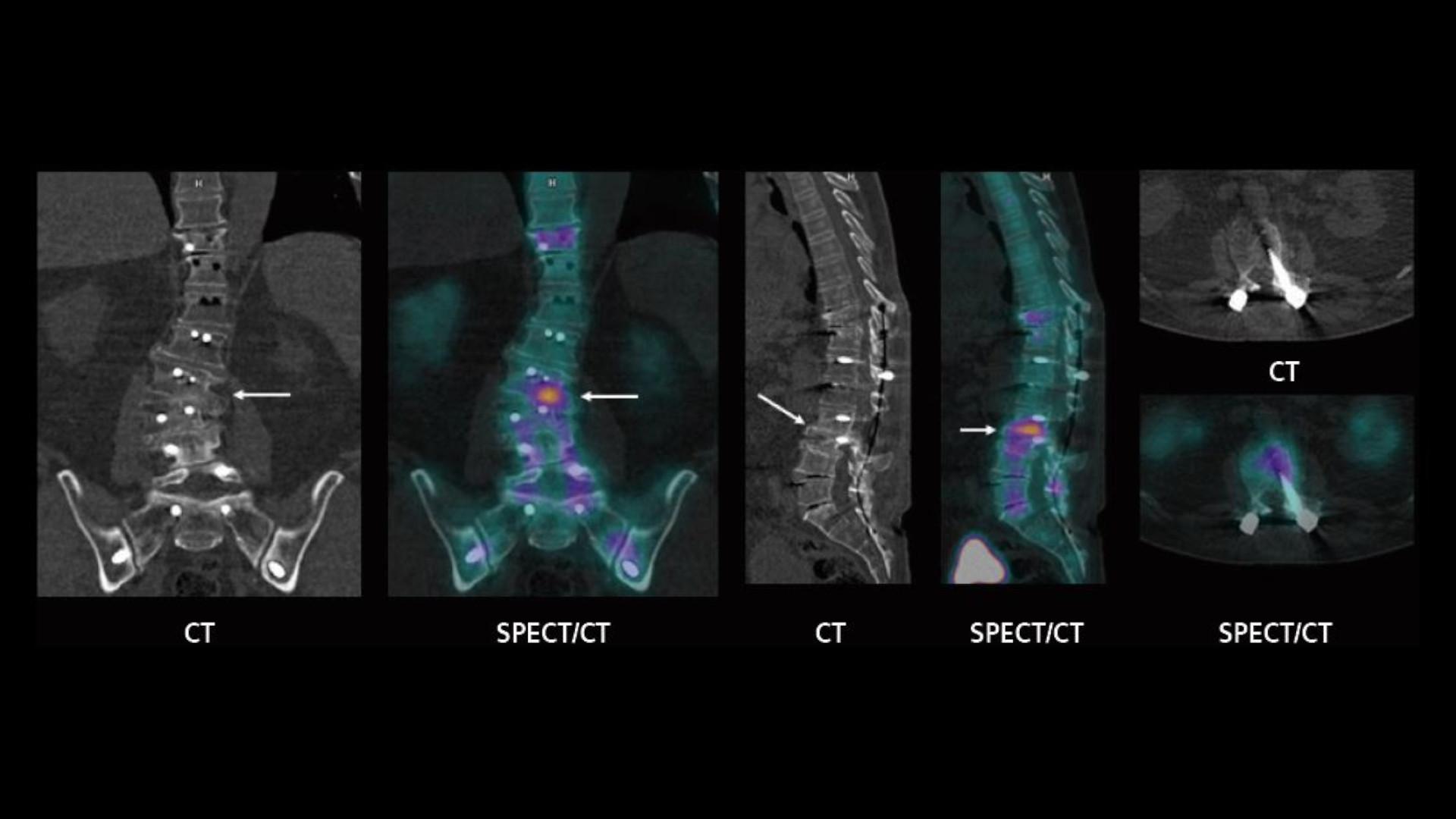
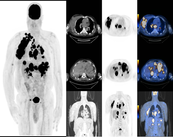
![Diagnostic performance of [68Ga]DOTATATE PET/CT, [18F]FDG PET/CT, MRI ...](https://cdn.ncbi.nlm.nih.gov/pmc/blobs/afe0/11399174/096009706f52/330_2024_10652_Fig3_HTML.jpg)


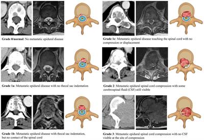
![Diagnostic performance of [68Ga]DOTATATE PET/CT, [18F]FDG PET/CT, MRI ...](https://cdn.ncbi.nlm.nih.gov/pmc/blobs/afe0/11399174/1c6e5e04e4e5/330_2024_10652_Fig4_HTML.jpg)


Enhance care with our medical Spinal Metastasis Pet/ct gallery of vast arrays of therapeutic images. clinically representing photography, images, and pictures. ideal for healthcare communications and materials. Discover high-resolution Spinal Metastasis Pet/ct images optimized for various applications. Suitable for various applications including web design, social media, personal projects, and digital content creation All Spinal Metastasis Pet/ct images are available in high resolution with professional-grade quality, optimized for both digital and print applications, and include comprehensive metadata for easy organization and usage. Our Spinal Metastasis Pet/ct gallery offers diverse visual resources to bring your ideas to life. Comprehensive tagging systems facilitate quick discovery of relevant Spinal Metastasis Pet/ct content. Whether for commercial projects or personal use, our Spinal Metastasis Pet/ct collection delivers consistent excellence. Reliable customer support ensures smooth experience throughout the Spinal Metastasis Pet/ct selection process. Our Spinal Metastasis Pet/ct database continuously expands with fresh, relevant content from skilled photographers. Each image in our Spinal Metastasis Pet/ct gallery undergoes rigorous quality assessment before inclusion. The Spinal Metastasis Pet/ct collection represents years of careful curation and professional standards. Multiple resolution options ensure optimal performance across different platforms and applications. Diverse style options within the Spinal Metastasis Pet/ct collection suit various aesthetic preferences.