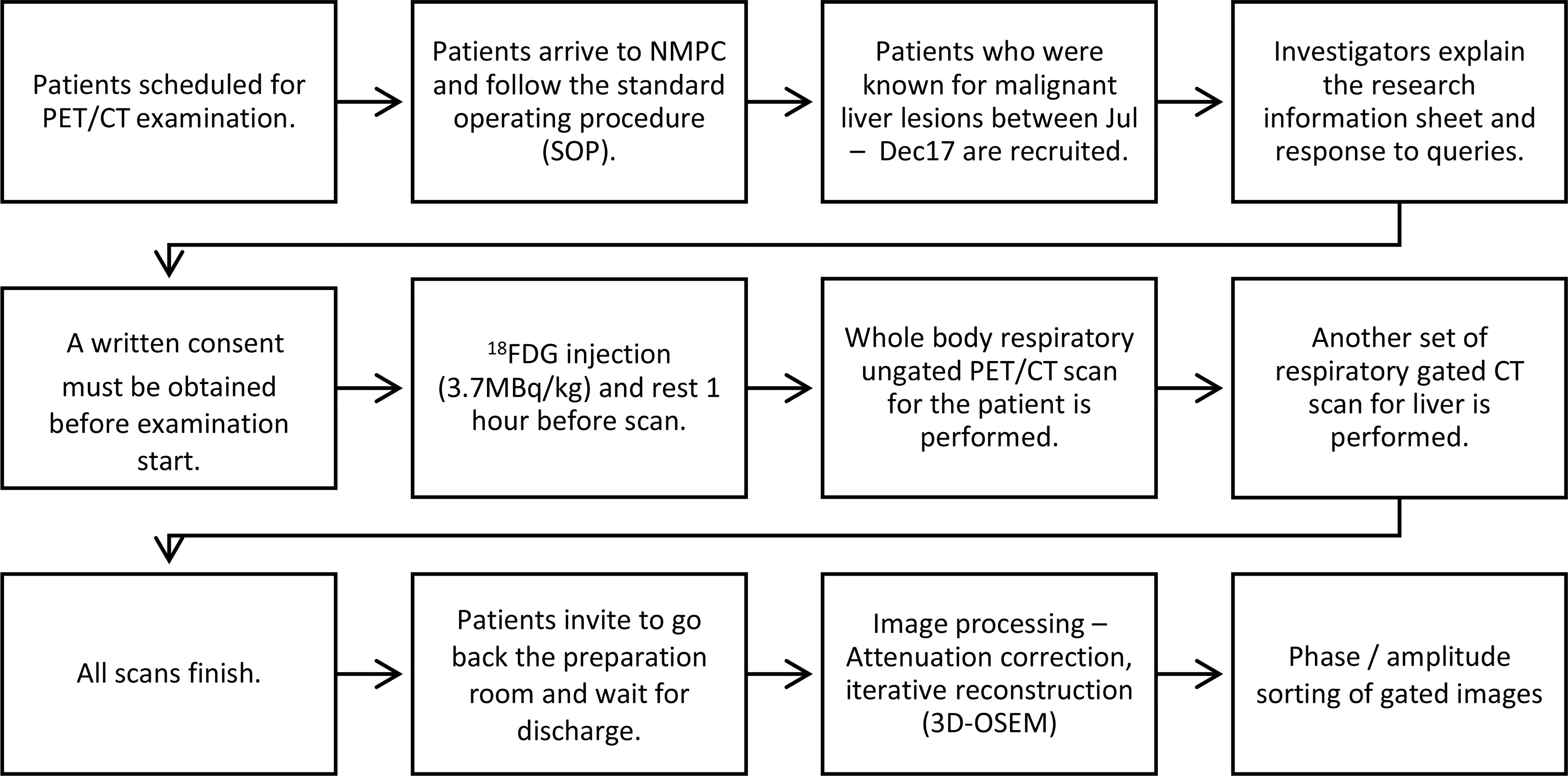
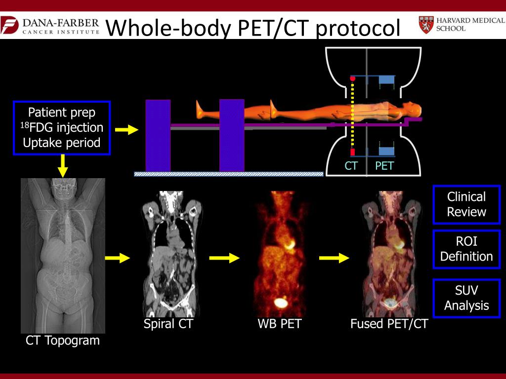
Pet/ct Workflow


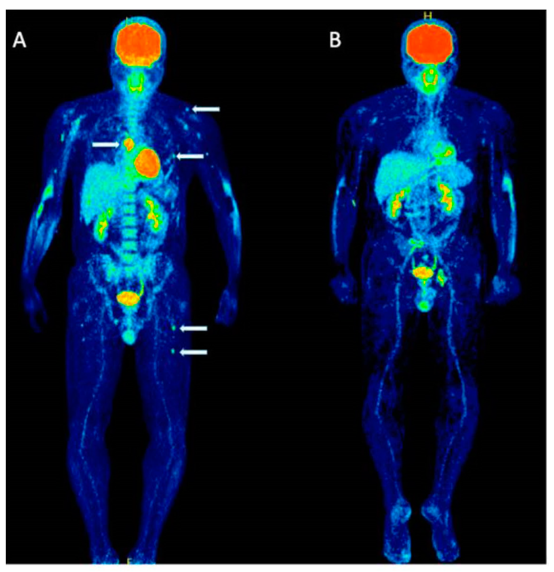
![(A,B transaxial fused images) manual workflow of [¹⁸F]F-choline PET/CT ...](https://www.researchgate.net/publication/364261414/figure/fig1/AS:11431281210472635@1702045801350/A-B-transaxial-fused-images-manual-workflow-of-FF-choline-PET-CT-imaging_Q640.jpg)


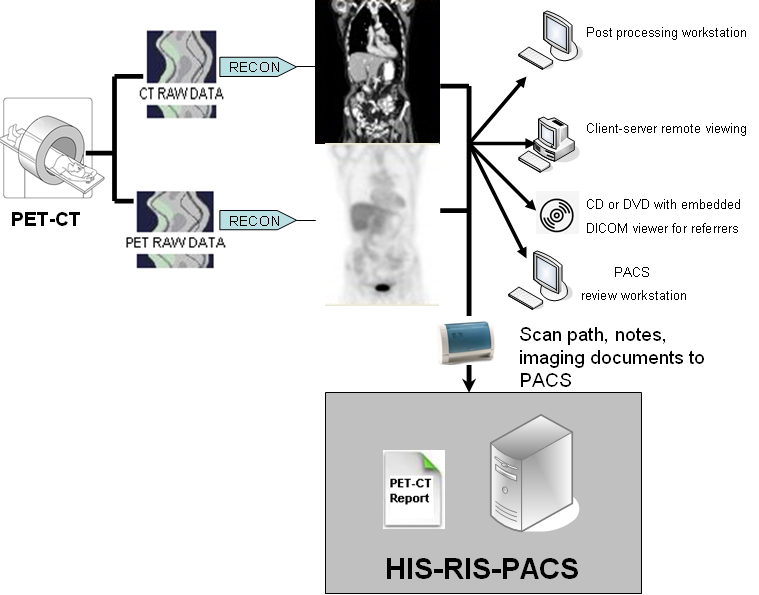
![[2409.09784] Enhancing Lesion Segmentation in PET/CT Imaging with Deep ...](https://ar5iv.labs.arxiv.org/html/2409.09784/assets/imgs/Workflow_dataCentric.png)


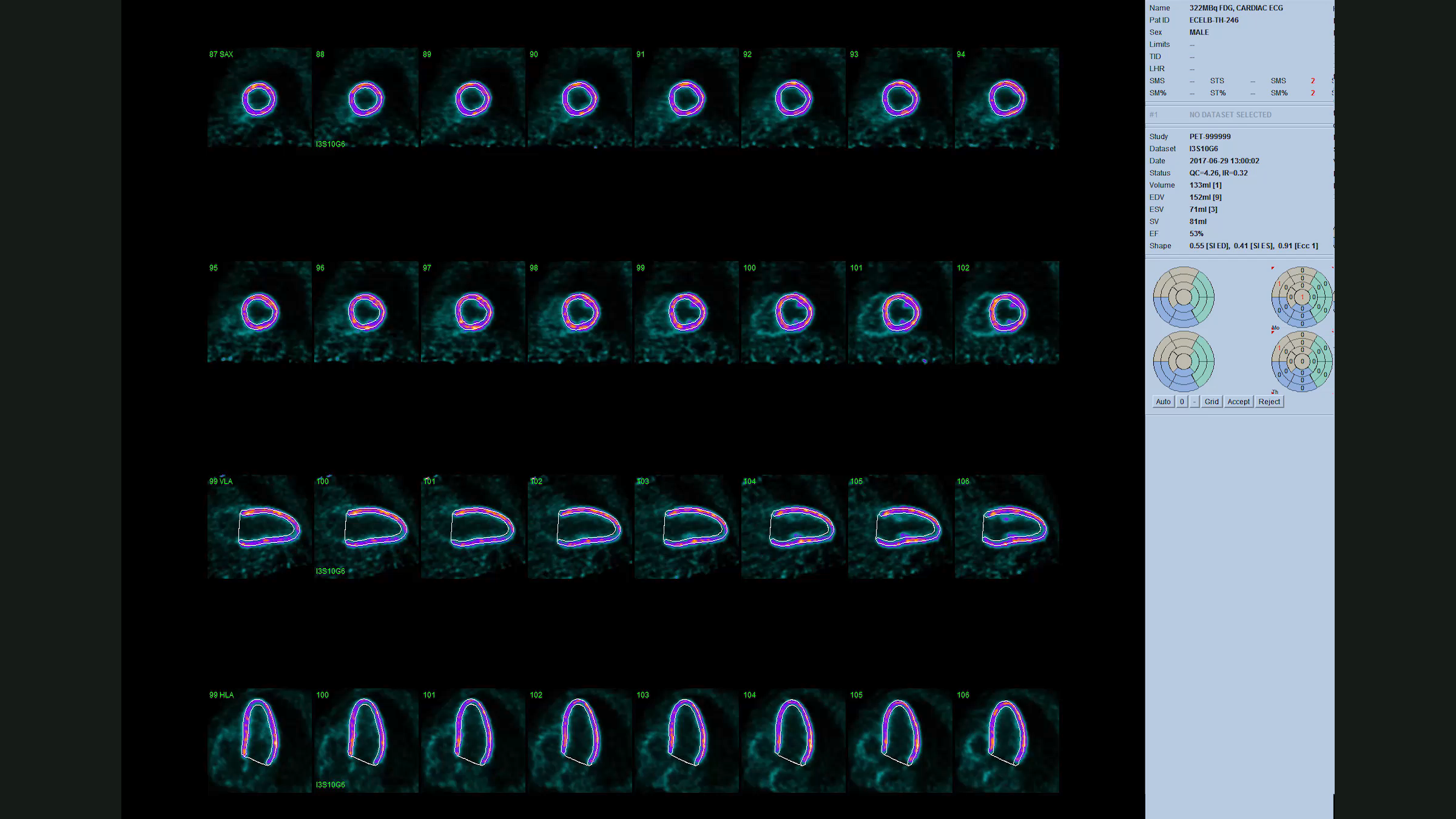


![Frontiers | Automated lesion detection of breast cancer in [18F] FDG ...](https://www.frontiersin.org/files/Articles/1007874/fonc-12-1007874-HTML/image_m/fonc-12-1007874-g001.jpg)


:max_bytes(150000):strip_icc()/2252467_color2-5bc4a212c9e77c00528a71be.png)


![Frontiers | Automated lesion detection of breast cancer in [18F] FDG ...](https://www.frontiersin.org/files/Articles/1007874/fonc-12-1007874-HTML/image_m/fonc-12-1007874-g004.jpg)


Study the characteristics of Pet/ct Workflow using our comprehensive set of extensive collections of learning images. enhancing knowledge retention through engaging and informative imagery. bridging theoretical knowledge with practical visual examples. Our Pet/ct Workflow collection features high-quality images with excellent detail and clarity. Excellent for educational materials, academic research, teaching resources, and learning activities All Pet/ct Workflow images are available in high resolution with professional-grade quality, optimized for both digital and print applications, and include comprehensive metadata for easy organization and usage. Our Pet/ct Workflow images support learning objectives across diverse educational environments. The Pet/ct Workflow archive serves professionals, educators, and creatives across diverse industries. Reliable customer support ensures smooth experience throughout the Pet/ct Workflow selection process. Our Pet/ct Workflow database continuously expands with fresh, relevant content from skilled photographers. Instant download capabilities enable immediate access to chosen Pet/ct Workflow images. The Pet/ct Workflow collection represents years of careful curation and professional standards. Regular updates keep the Pet/ct Workflow collection current with contemporary trends and styles. Diverse style options within the Pet/ct Workflow collection suit various aesthetic preferences. Multiple resolution options ensure optimal performance across different platforms and applications.