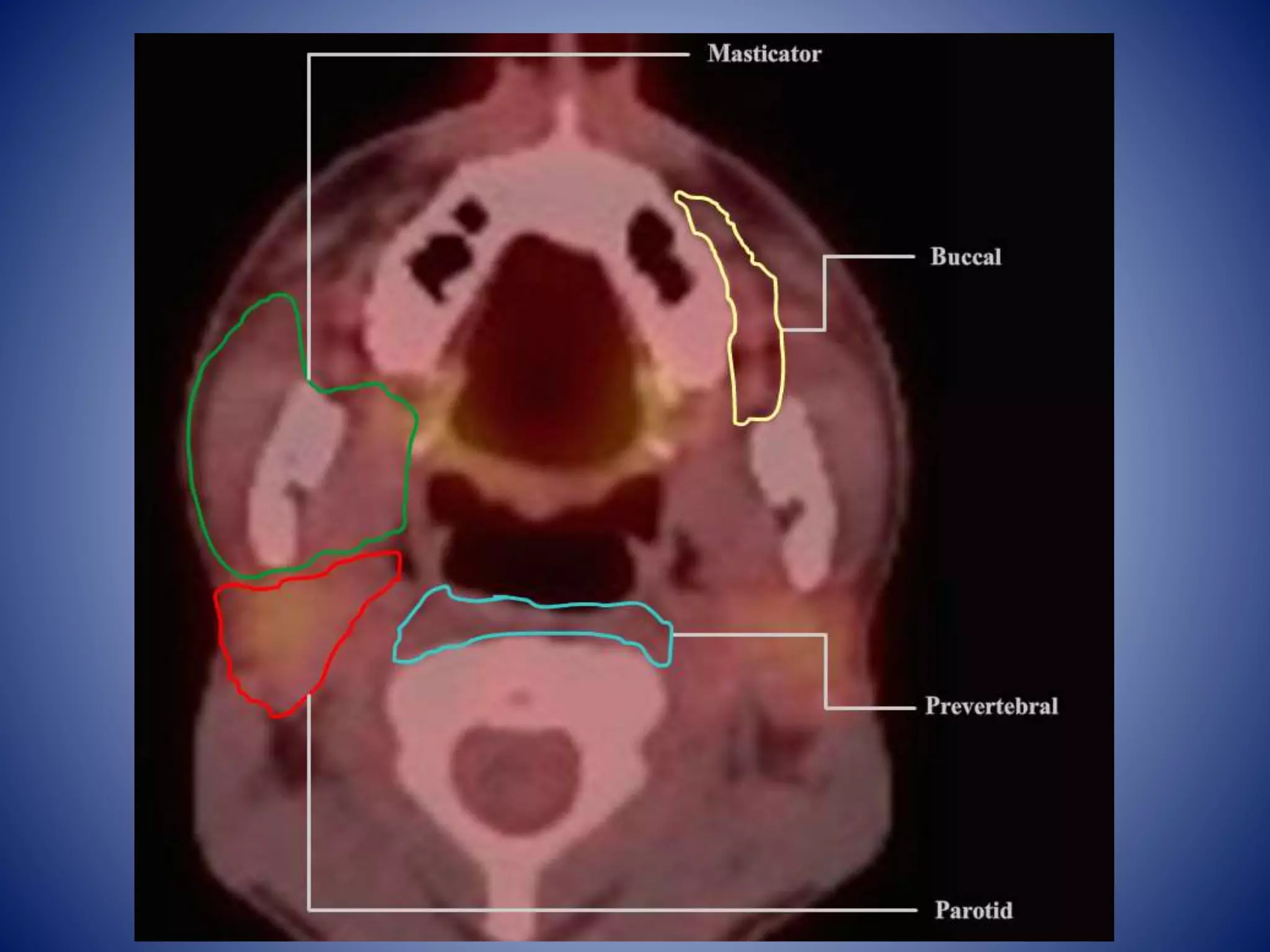
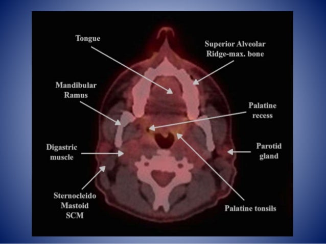
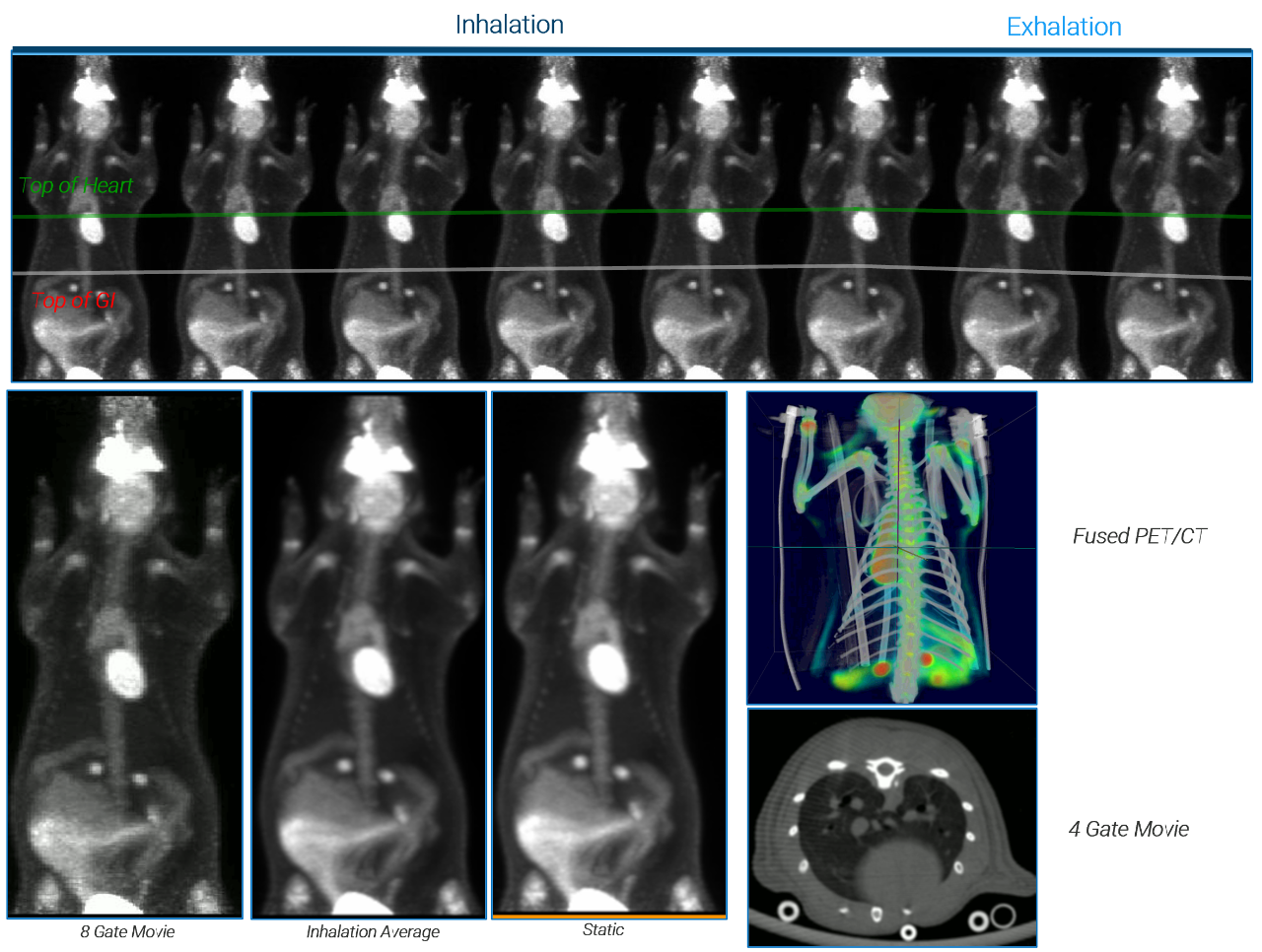
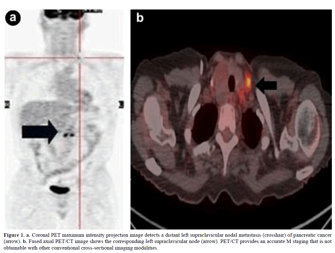
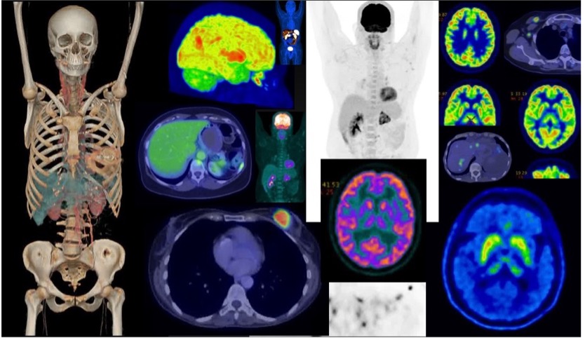
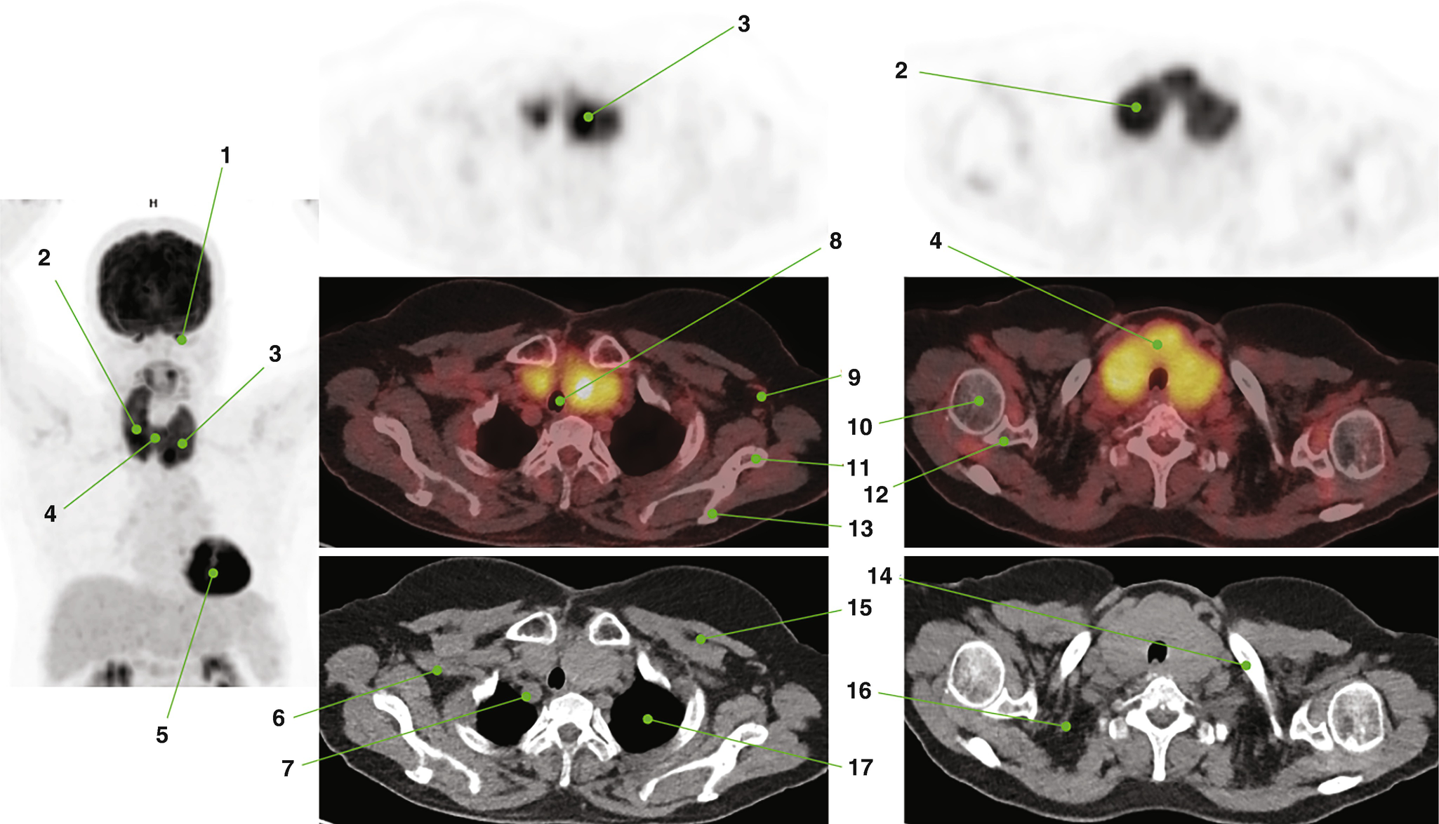
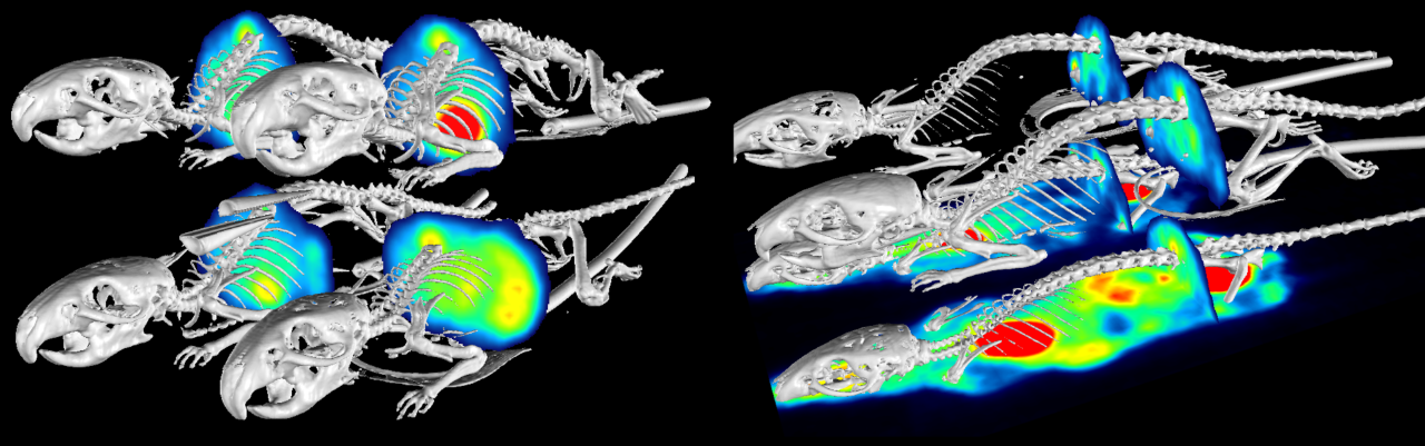
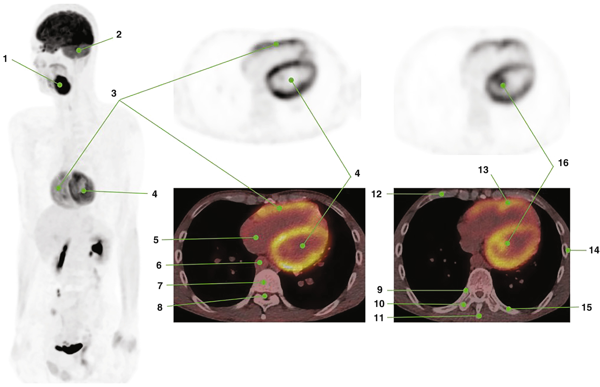
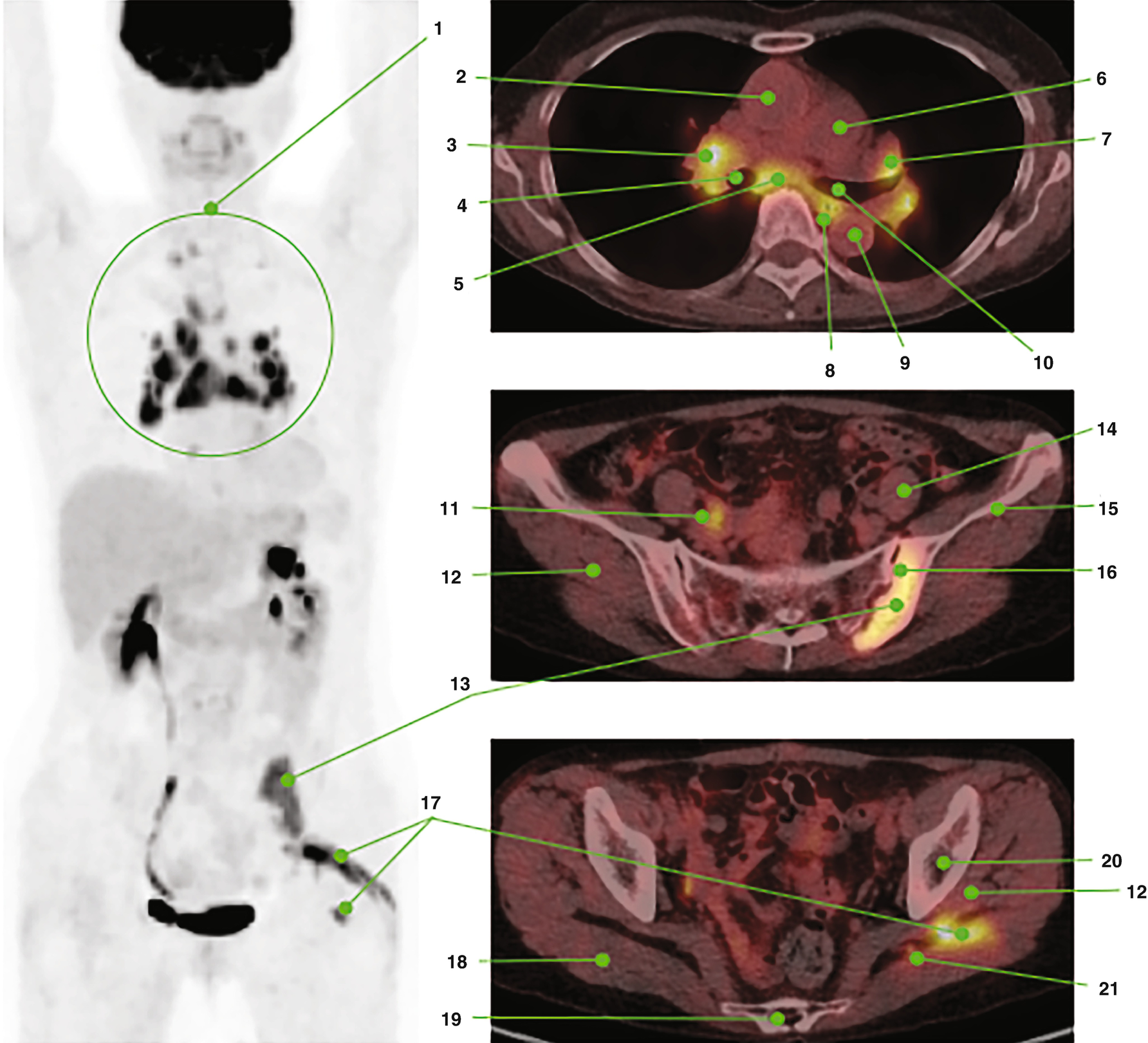
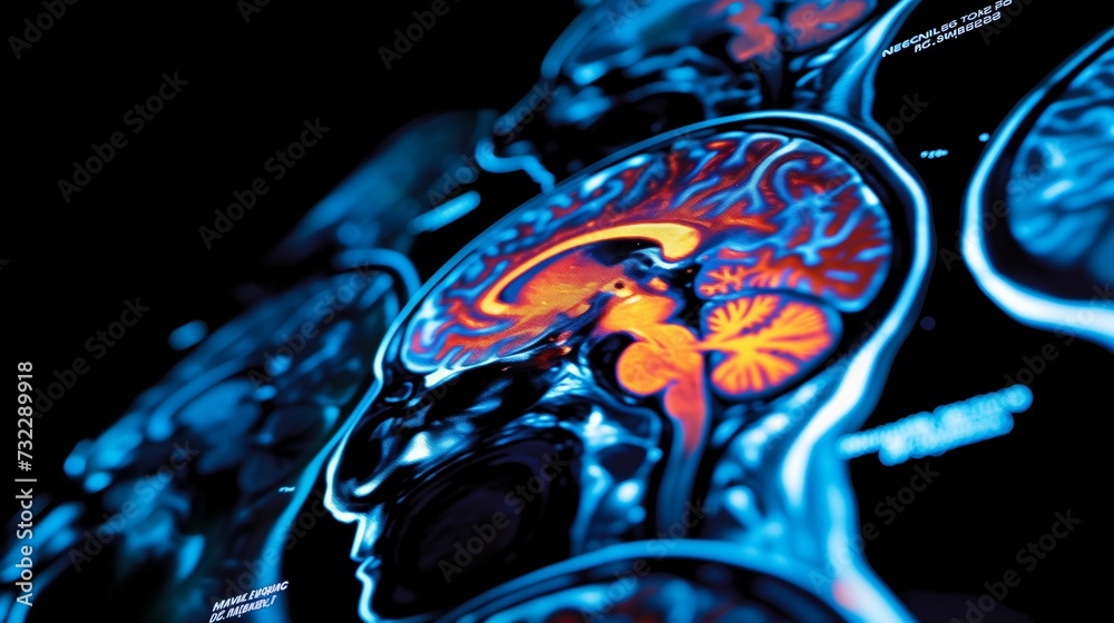
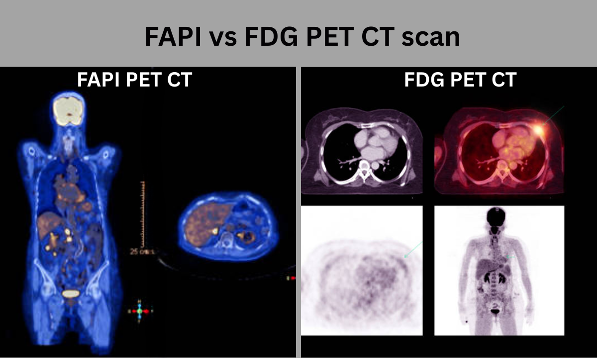
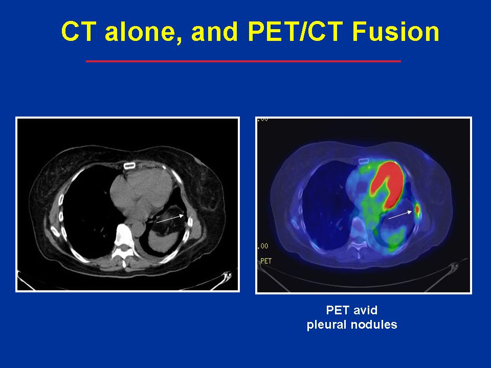
Pet/ct Sectional Images


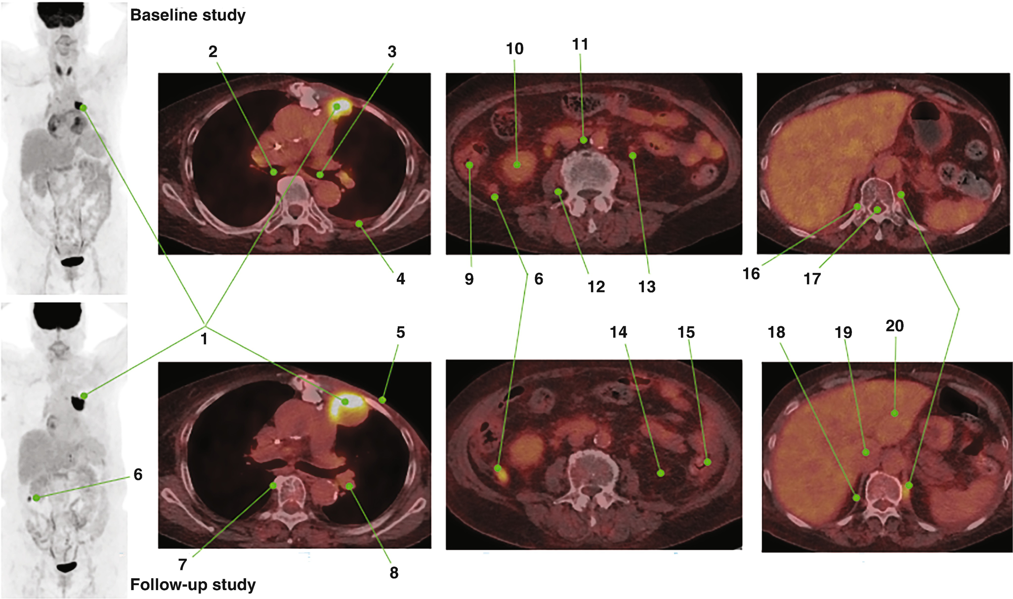


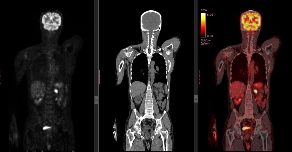
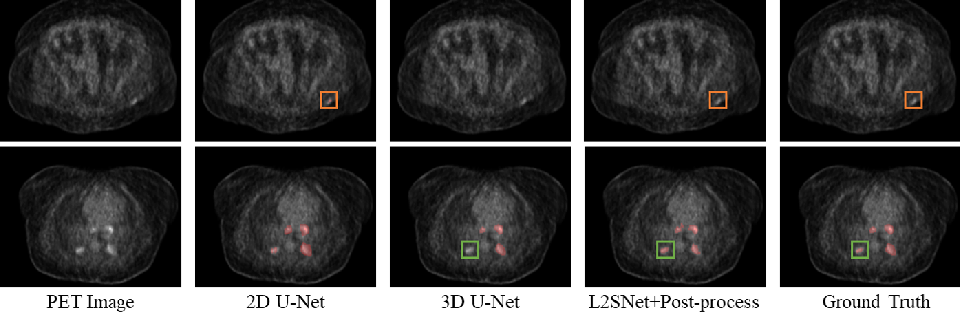
![Figure 1, [18F-FDG PET/CT imaging. Baseline whole...]. - Molecular ...](https://www.ncbi.nlm.nih.gov/books/NBK599131/bin/chapter2_f1.jpg)


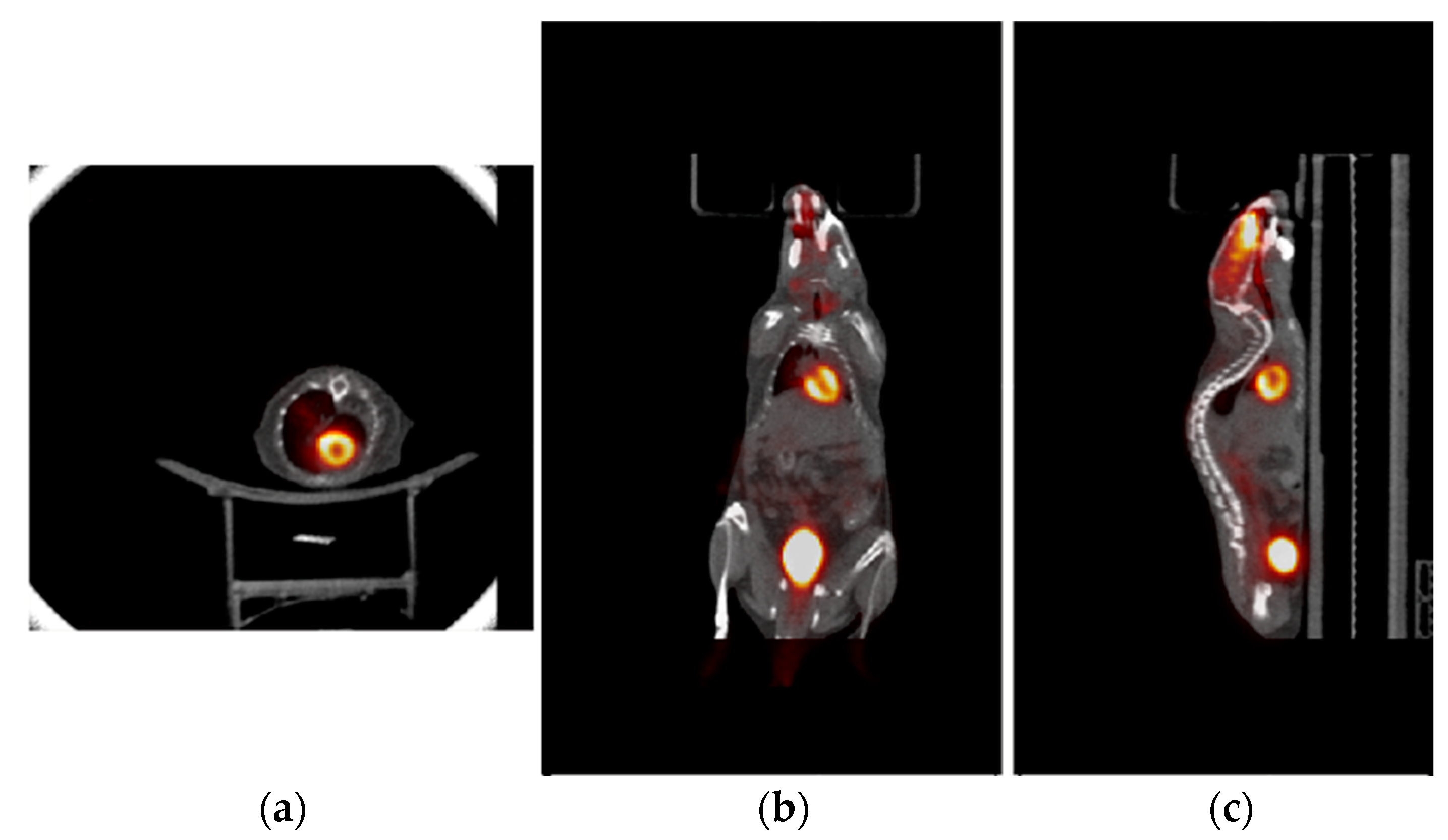


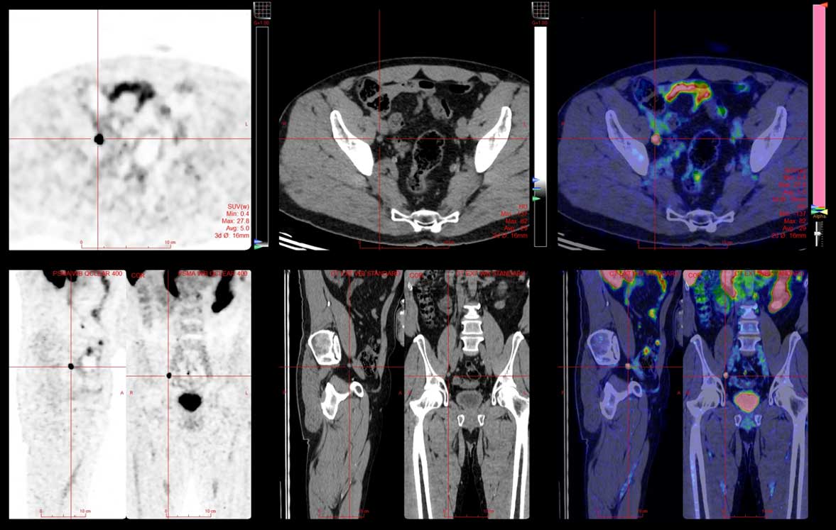
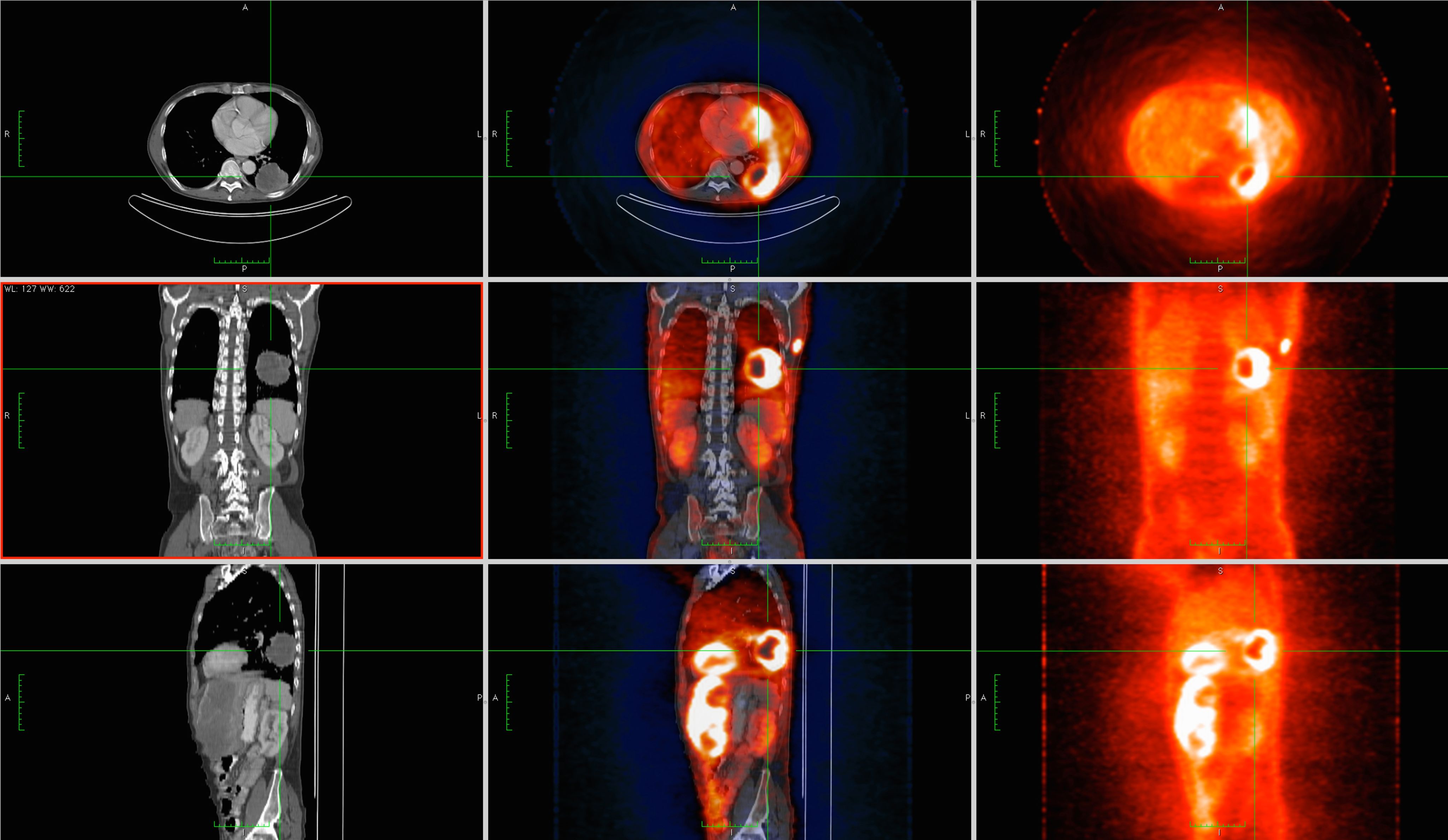
![-PET [right] and PET-CT fusion images [left] axial sections showing the ...](https://www.researchgate.net/publication/351299512/figure/fig3/AS:1025582590611456@1621529352559/PET-right-and-PET-CT-fusion-images-left-axial-sections-showing-the-increased-uptake.jpg)


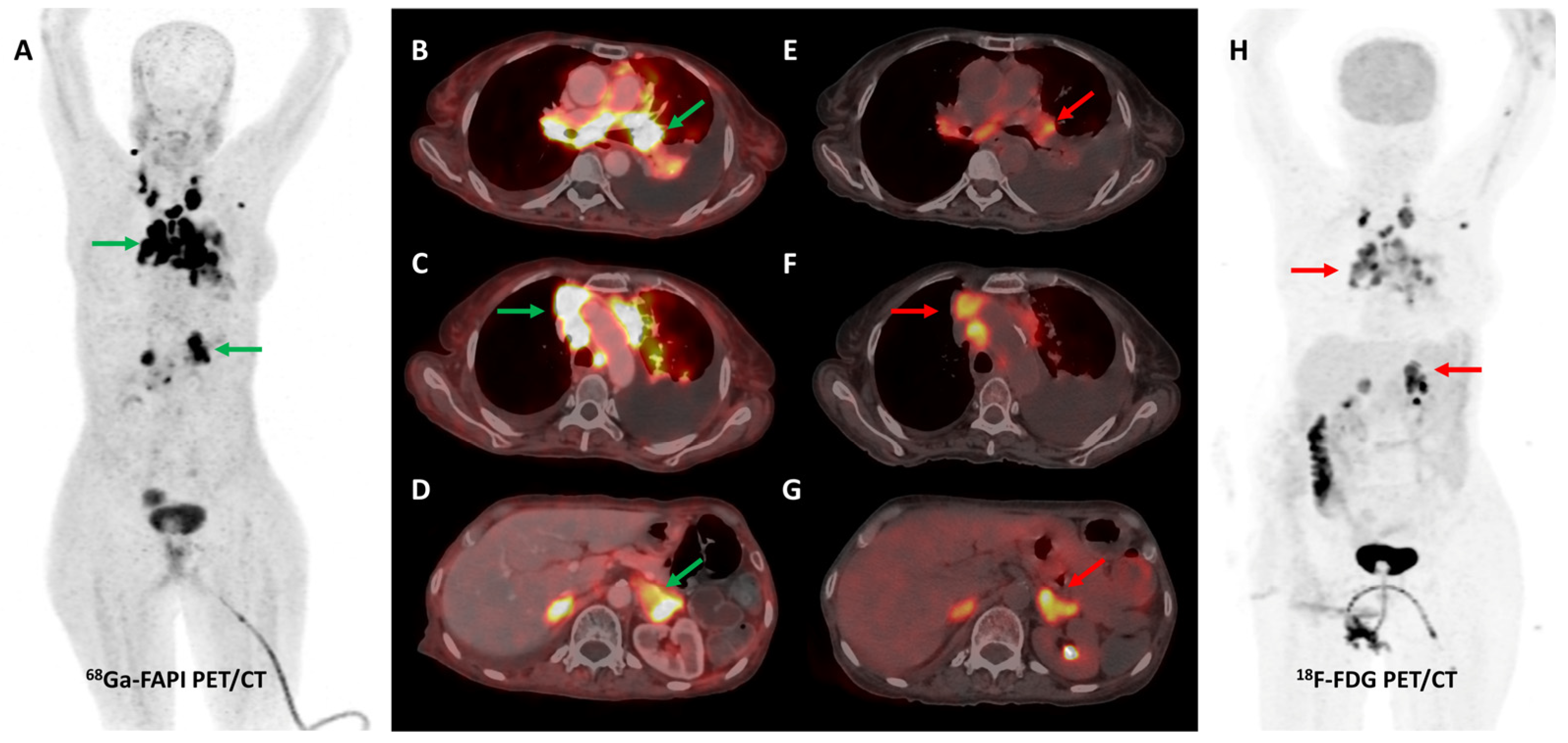
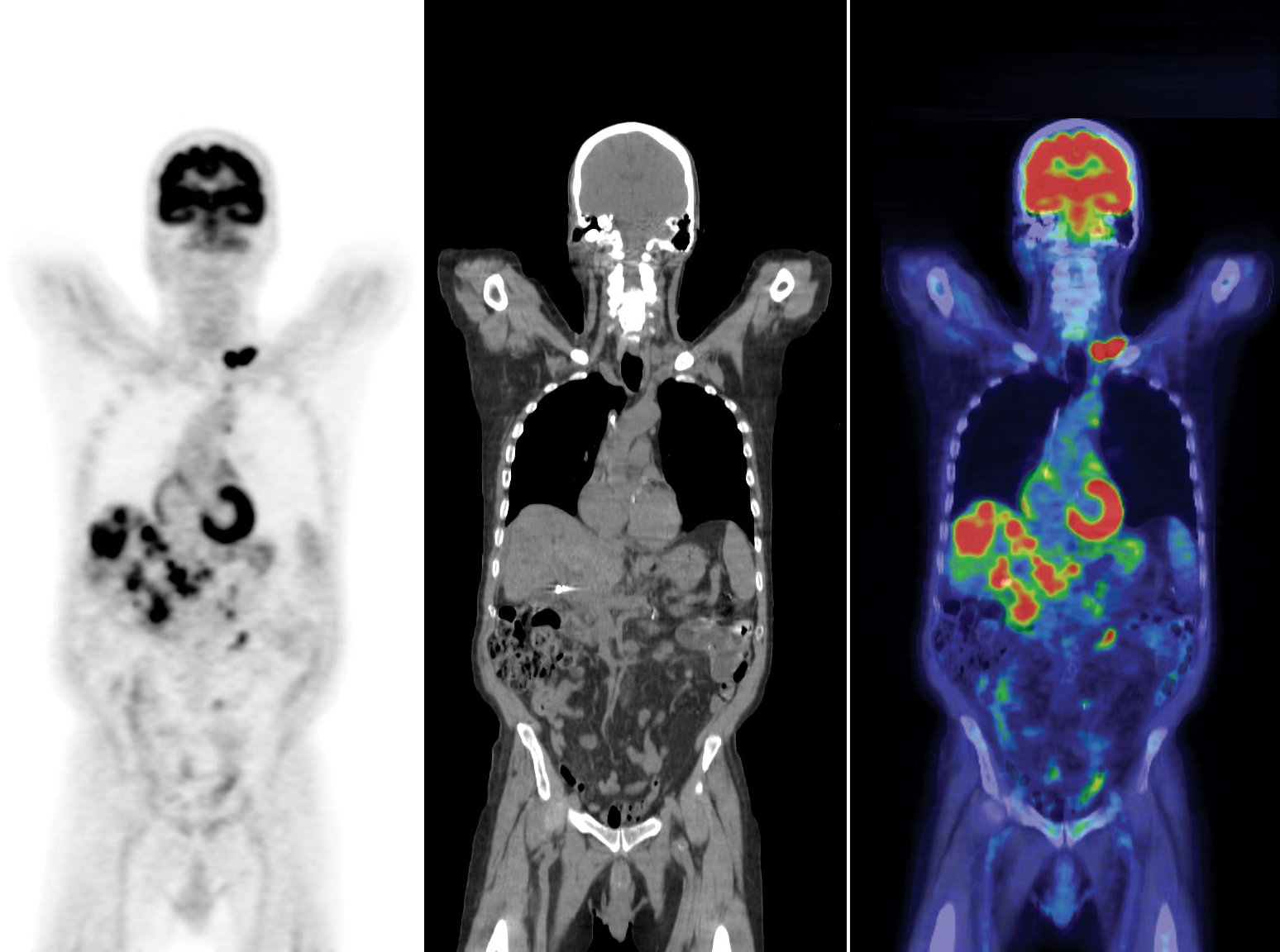
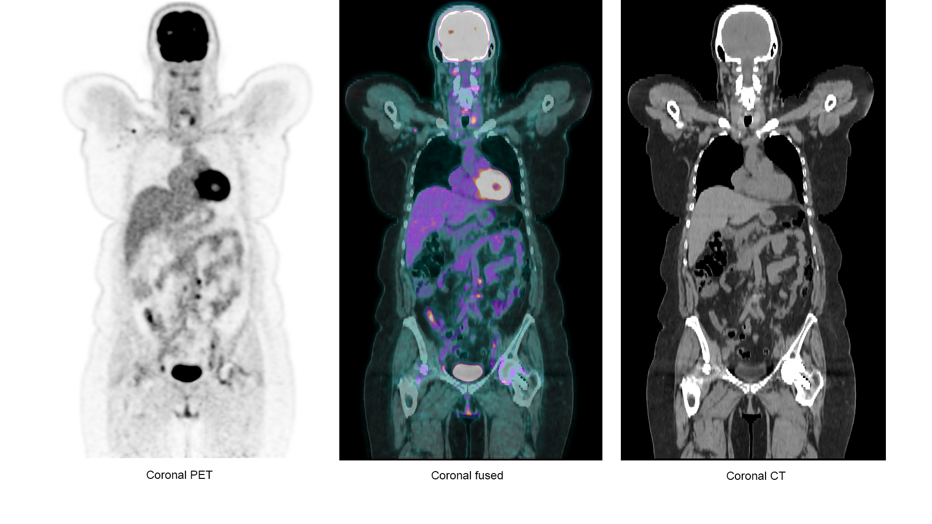
![Axial section of the [ 18 F]FDG-PET/CT scan of patient A (A-D) and ...](https://www.researchgate.net/publication/365148559/figure/fig1/AS:11431281095108384@1667745105476/Axial-section-of-the-18-FFDG-PET-CT-scan-of-patient-A-A-D-and-patient-B-E-H-before_Q320.jpg)


:max_bytes(150000):strip_icc()/VWH-GettyImages-1876736668-f317c729fd0b4e5eb264ae5add38b7bb.jpg)


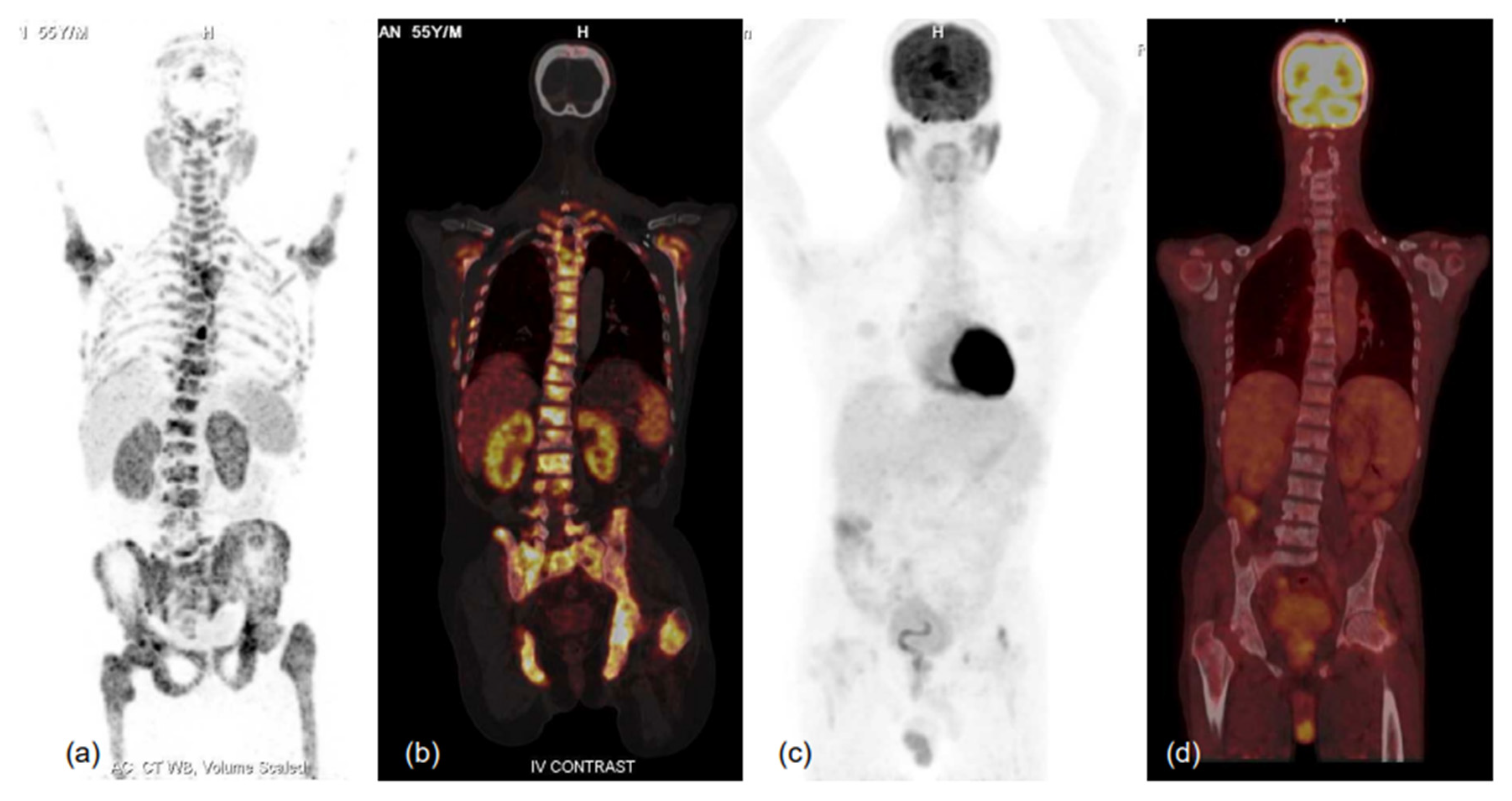
![[ 18 F]FDG PET-CT scan. Axial section showing diffusely increased ...](https://www.researchgate.net/profile/Awadhesh-Tiwari-4/publication/373643364/figure/fig1/AS:11431281185972496@1693810656380/18-FFDG-PET-CT-scan-Axial-section-showing-diffusely-increased-metabolic-activity-in_Q320.jpg)


Design the future through extensive collections of architecture-focused Pet/ct Sectional Images photographs. architecturally showcasing photography, pictures, and visuals. designed to inspire architectural innovation. Browse our premium Pet/ct Sectional Images gallery featuring professionally curated photographs. Suitable for various applications including web design, social media, personal projects, and digital content creation All Pet/ct Sectional Images are available in high resolution with professional-grade quality, optimized for both digital and print applications, and include comprehensive metadata for easy organization and usage. Discover the perfect Pet/ct Sectional Images to enhance your visual communication needs. Reliable customer support ensures smooth experience throughout the Pet/ct Sectional Images selection process. Comprehensive tagging systems facilitate quick discovery of relevant Pet/ct Sectional Images content. The Pet/ct Sectional Images archive serves professionals, educators, and creatives across diverse industries. Multiple resolution options ensure optimal performance across different platforms and applications. The Pet/ct Sectional Images collection represents years of careful curation and professional standards. Regular updates keep the Pet/ct Sectional Images collection current with contemporary trends and styles. Instant download capabilities enable immediate access to chosen Pet/ct Sectional Images images. Professional licensing options accommodate both commercial and educational usage requirements. Advanced search capabilities make finding the perfect Pet/ct Sectional Images image effortless and efficient.