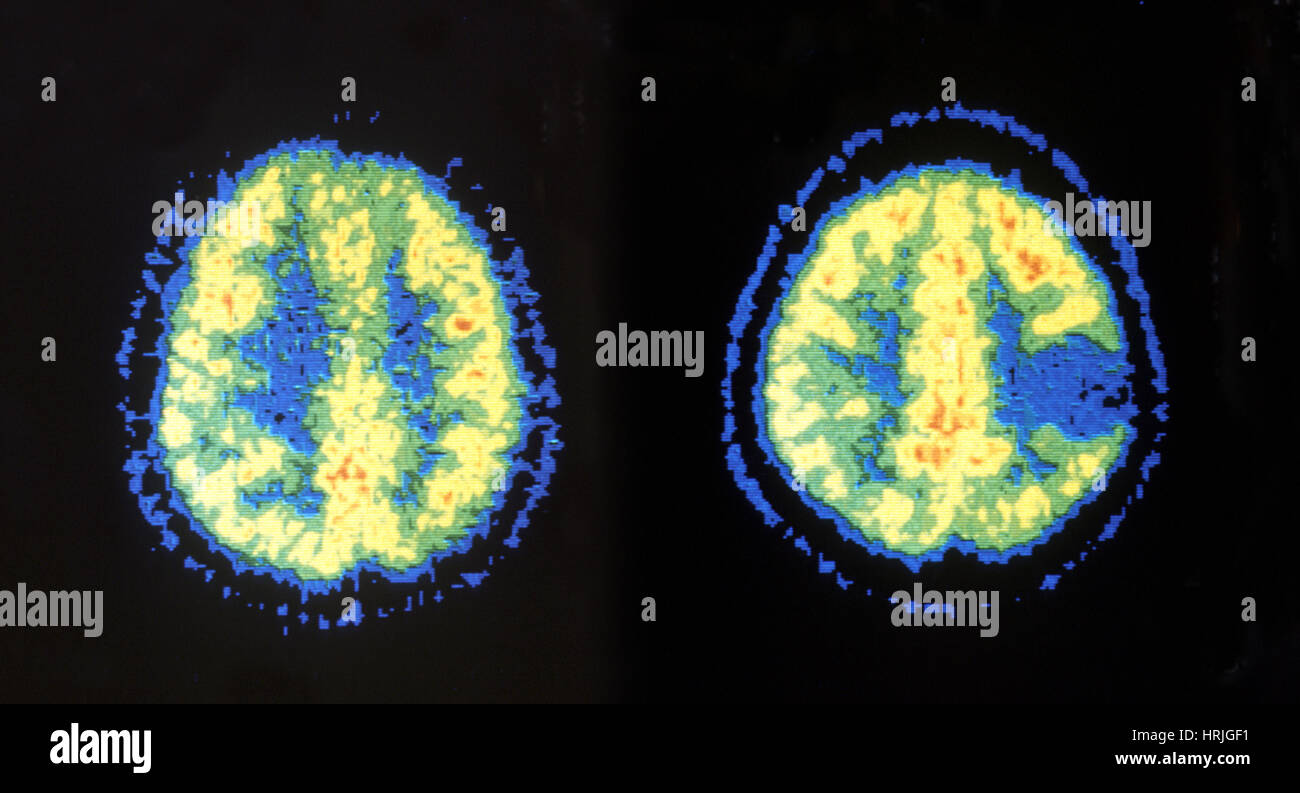
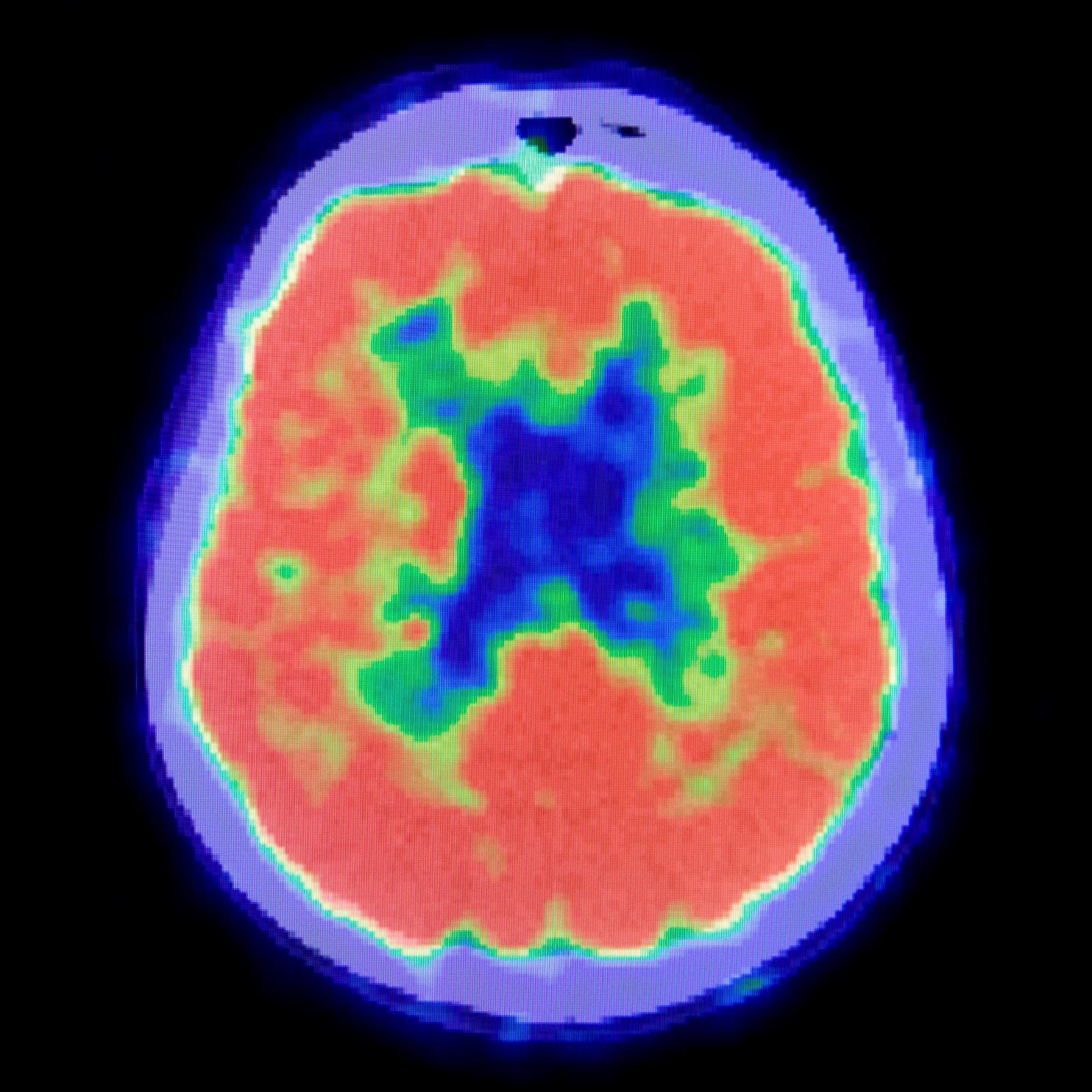
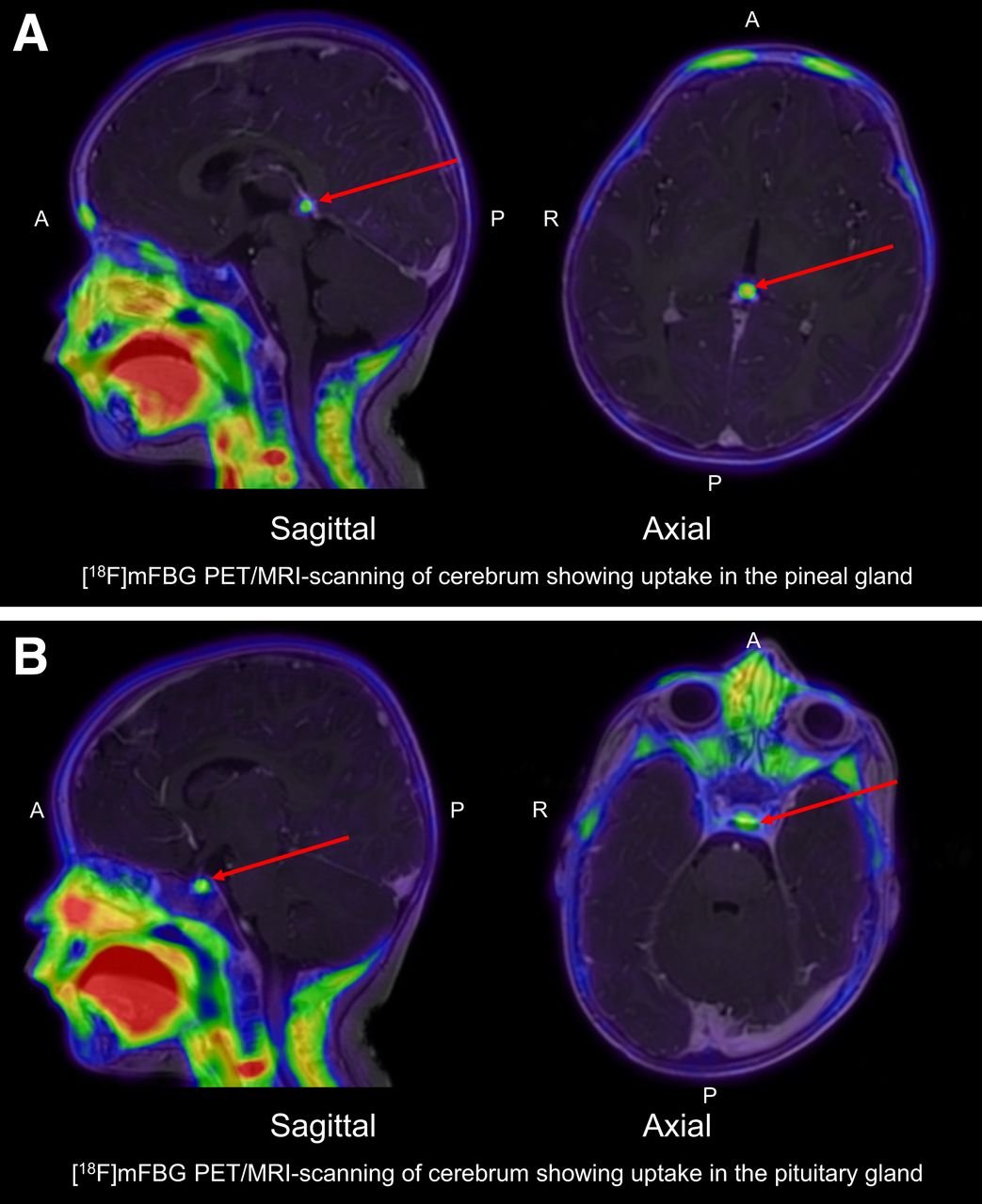
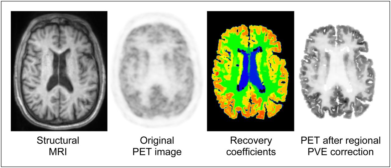
Normal Pet/ct Brain


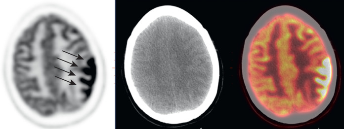


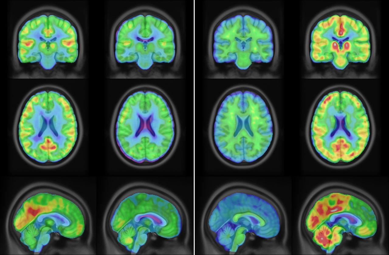
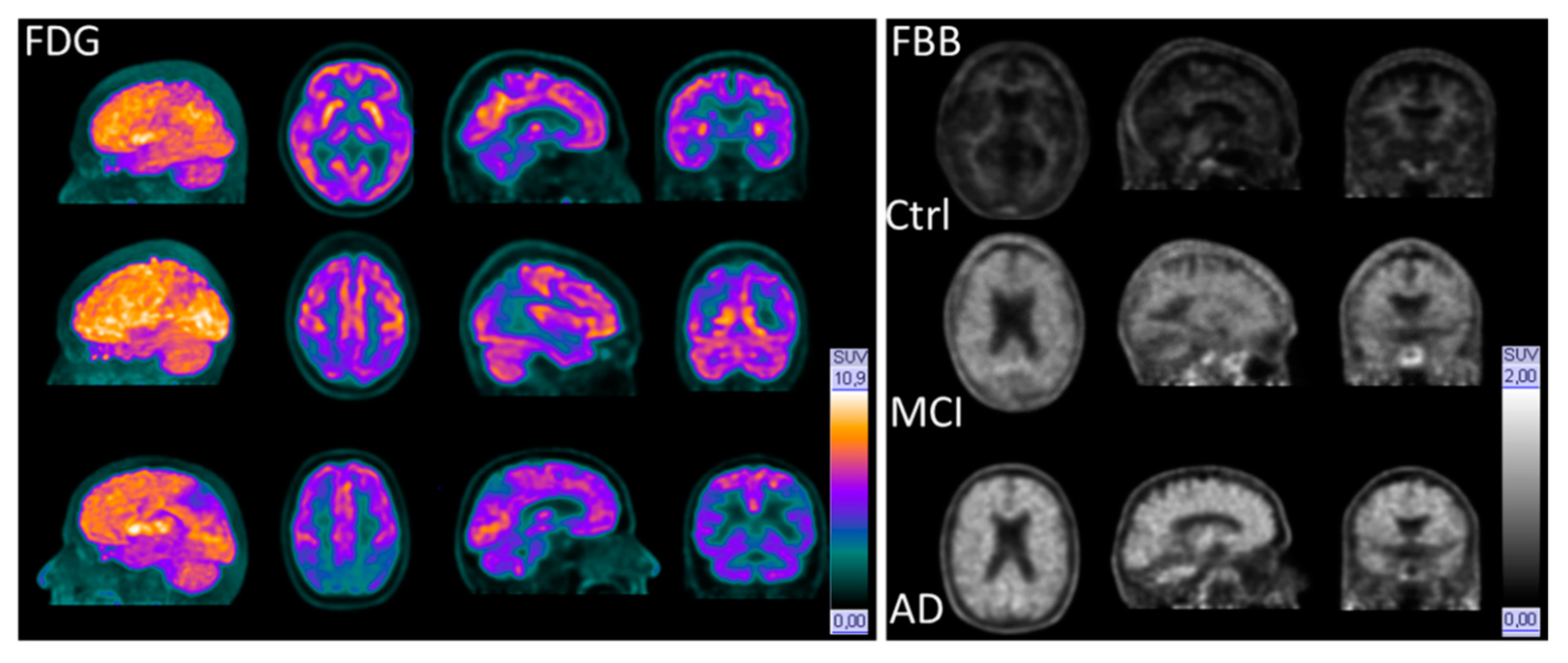
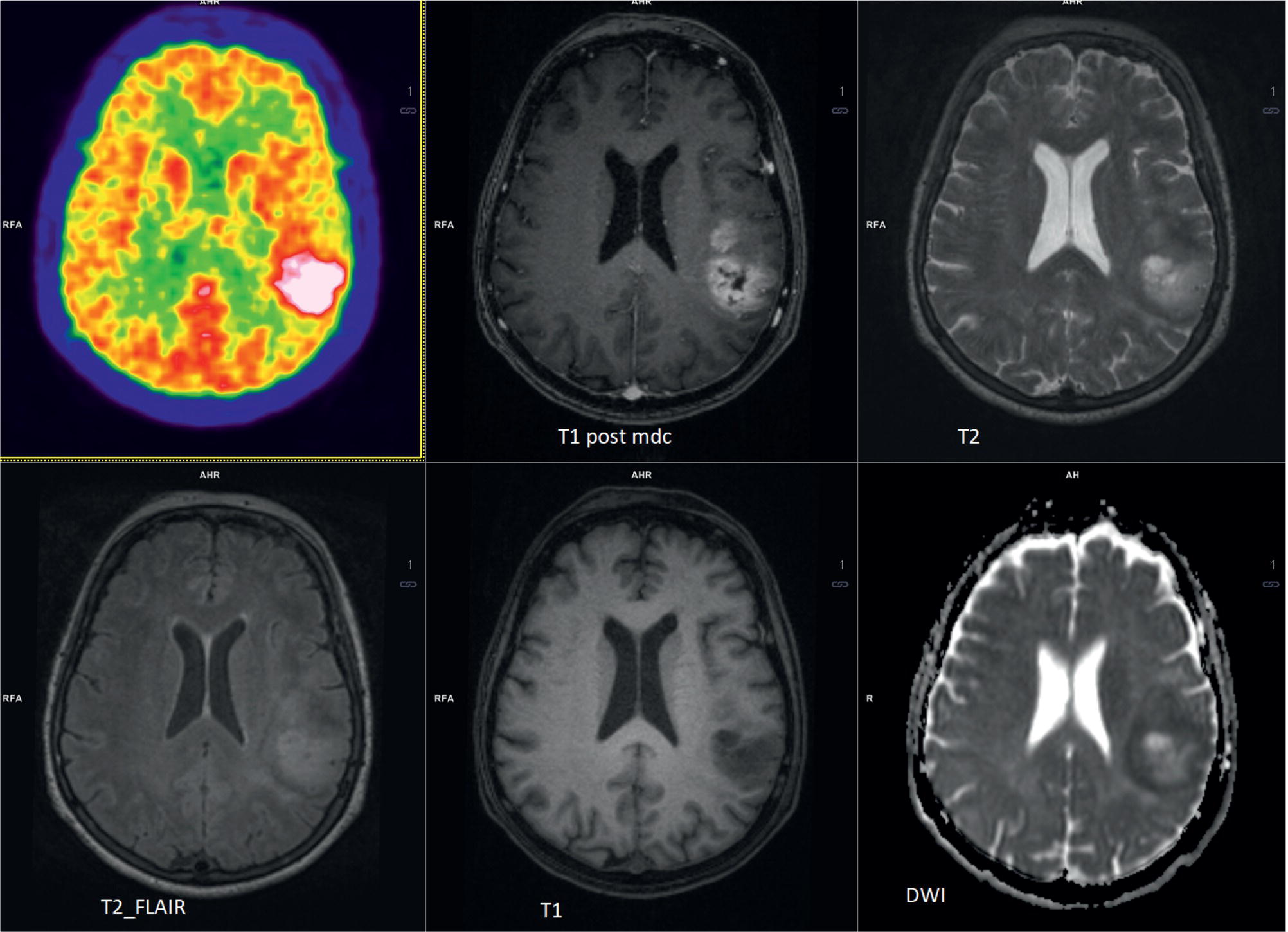
![[Figure, FDG PET/CT Brain Scan Revealing...] - StatPearls - NCBI Bookshelf](https://www.ncbi.nlm.nih.gov/books/NBK603715/bin/Breast__Cancer__Patient__FDG__PET__Brain__Images__with__Two__Different__PET__Windows_2.jpg)


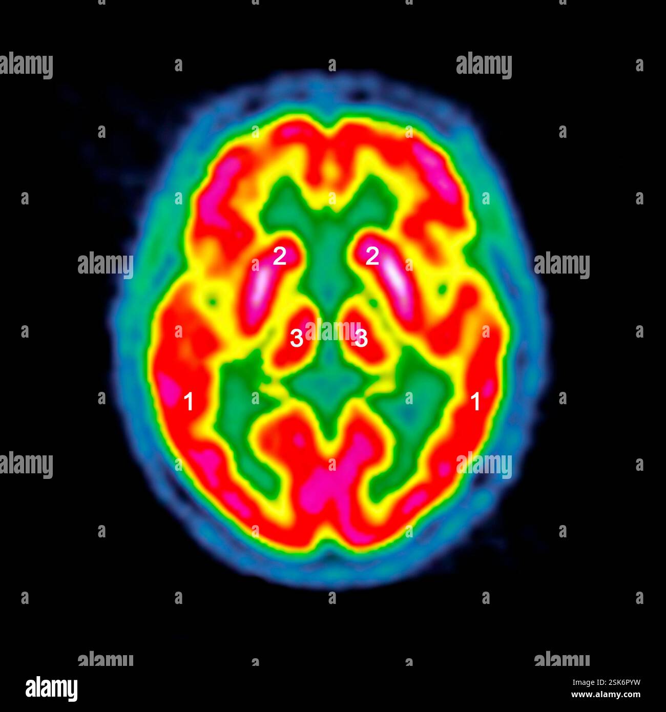
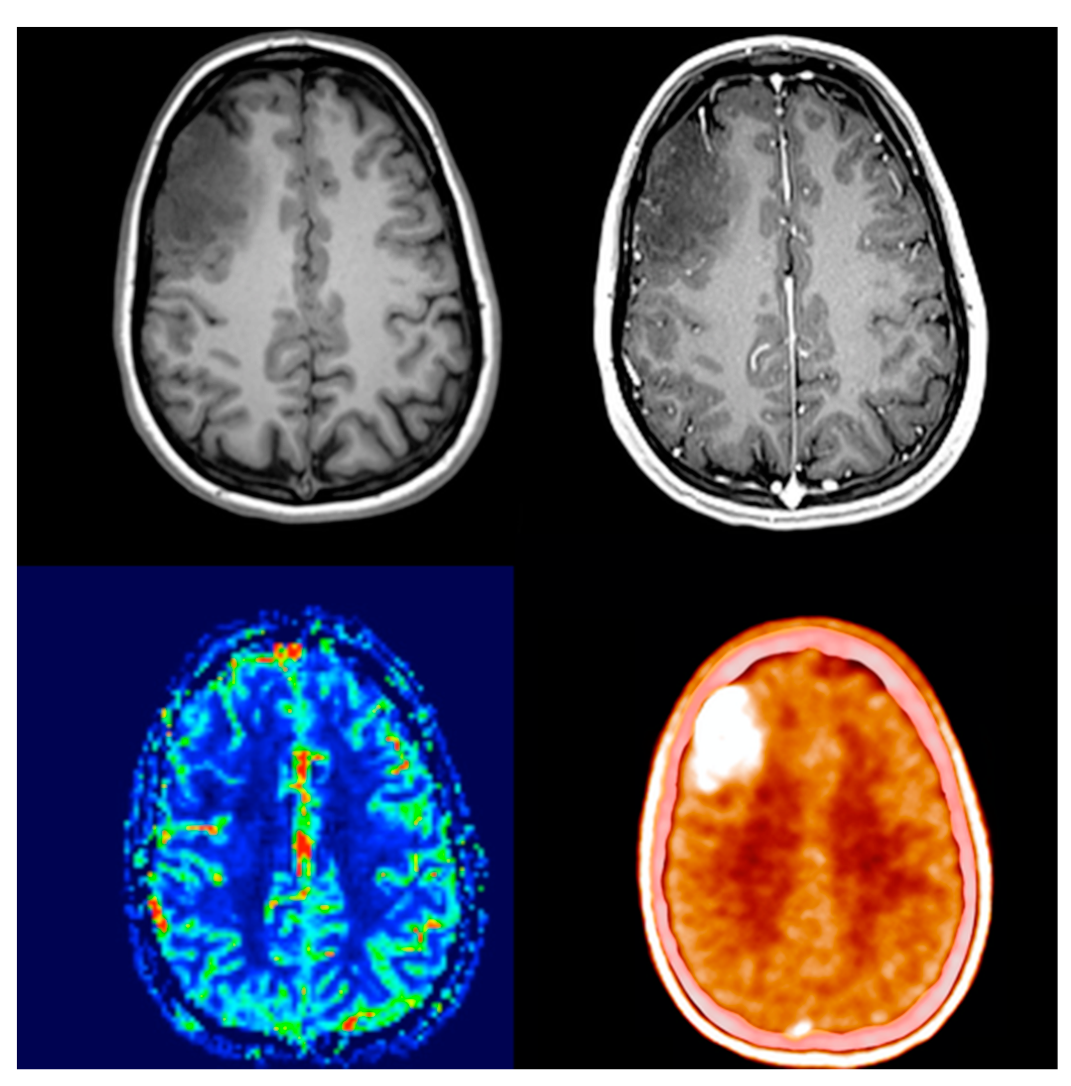
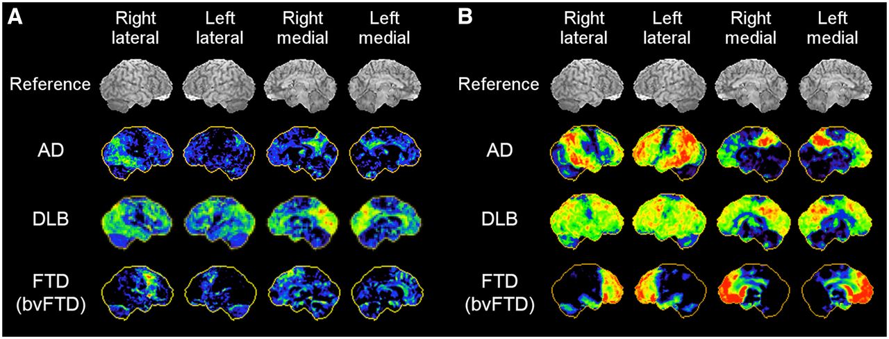
![[Figure, F18 FDG PET/CT Brain Scan...] - StatPearls - NCBI Bookshelf](https://www.ncbi.nlm.nih.gov/books/NBK603715/bin/DLB1.jpg)


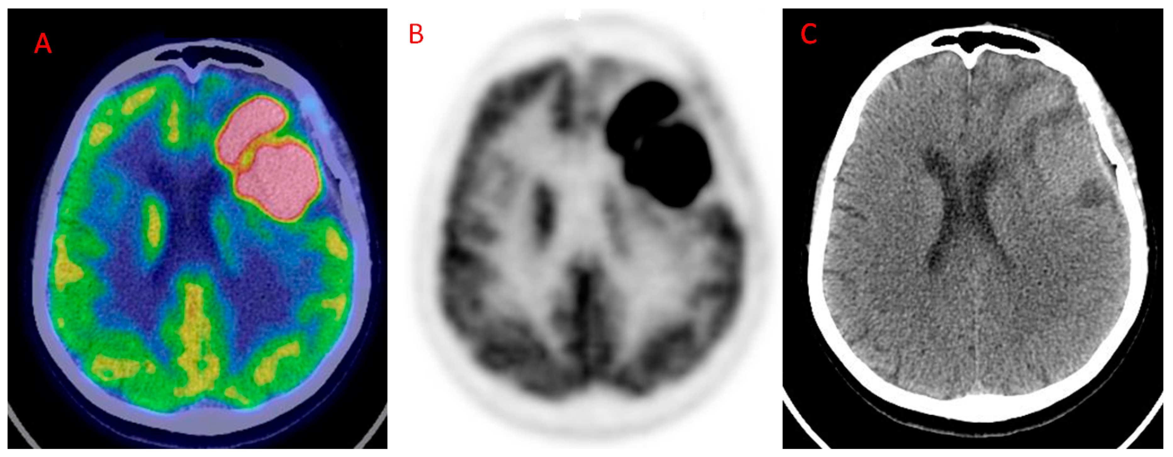
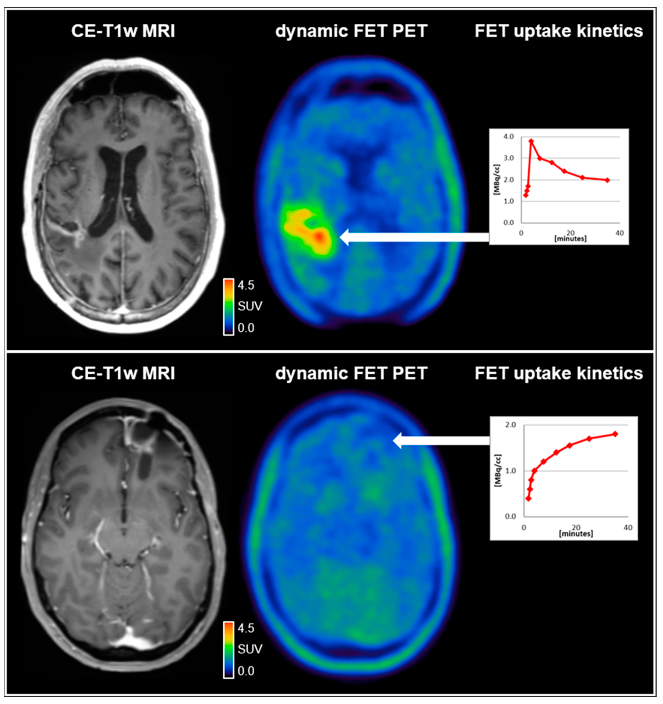
![A, B: Brain PET/CT imaging with [ 18 F]FET in a patient with suspected ...](https://www.researchgate.net/publication/296693886/figure/fig5/AS:335582011117578@1457020394848/A-B-Brain-PET-CT-imaging-with-18-FFET-in-a-patient-with-suspected-recurrence-of.png)
![Representative horizontal brain PET/CT images of [ 18 F]3 d 7 in WT and ...](https://www.researchgate.net/publication/326205339/figure/fig1/AS:871599112724484@1584816830503/Representative-horizontal-brain-PET-CT-images-of-18-F3-d-7-in-WT-and-Mdr1a-b.png)


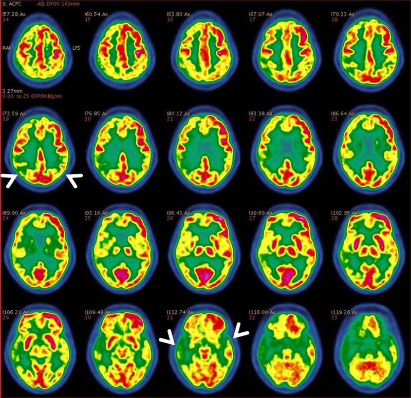

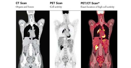


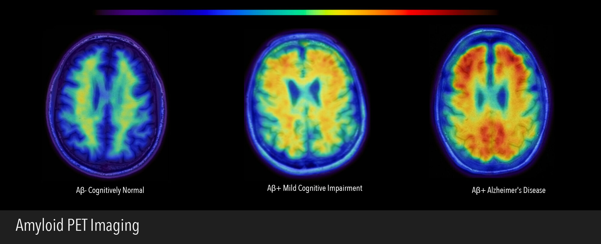


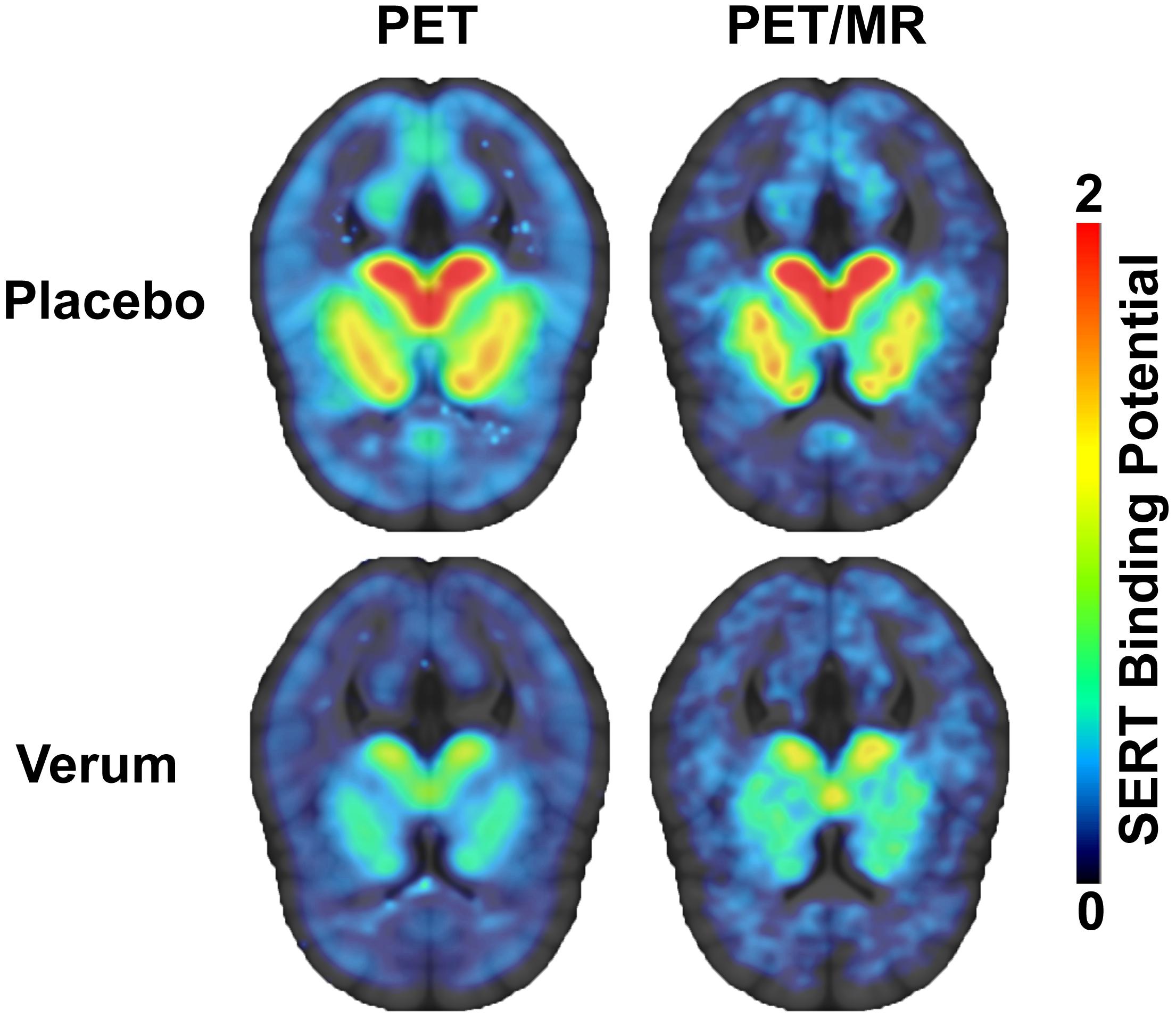

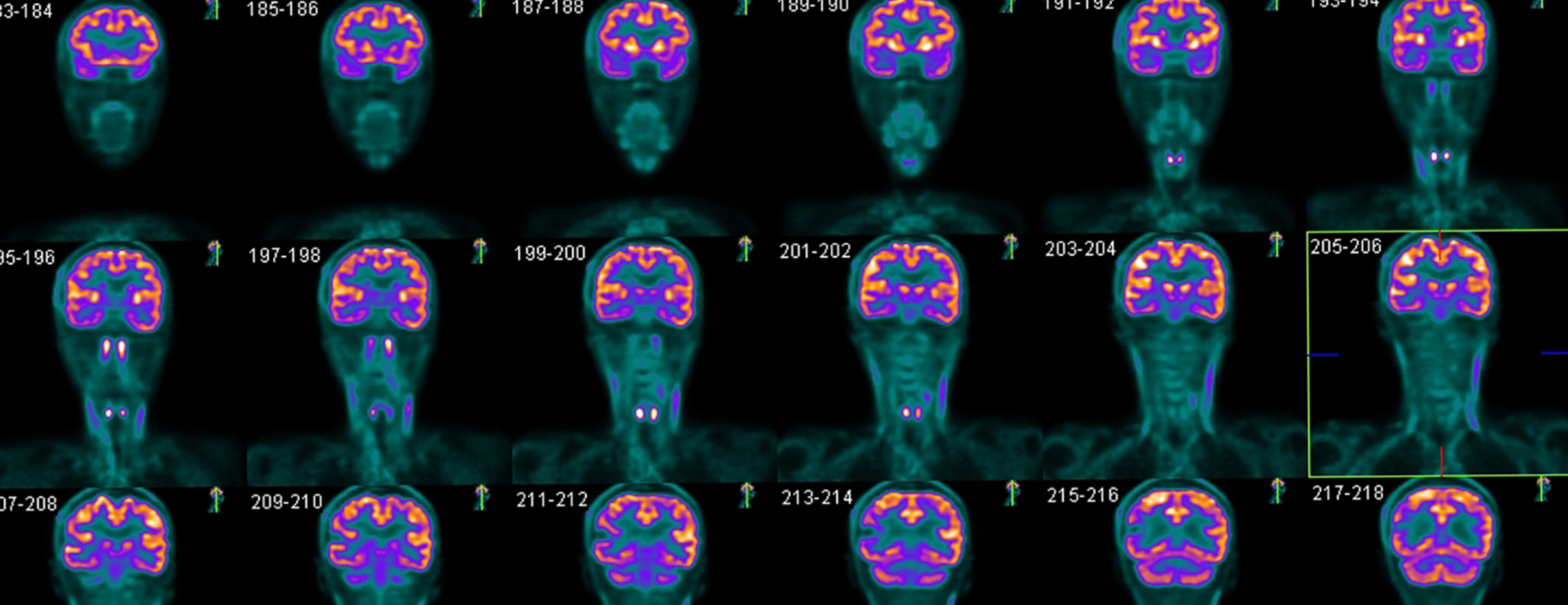
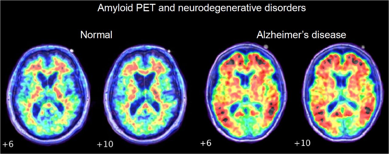


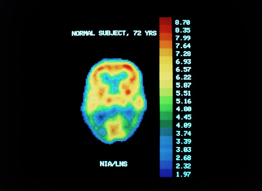
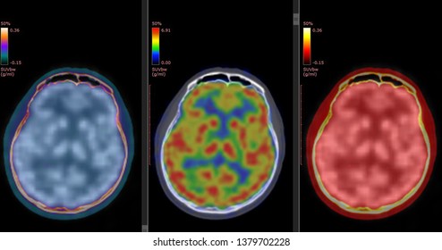


Discover traditional Normal Pet/ct Brain with our collection of hundreds of classic photographs. preserving the heritage of computer, digital, and electronic. designed to preserve cultural significance. Browse our premium Normal Pet/ct Brain gallery featuring professionally curated photographs. Suitable for various applications including web design, social media, personal projects, and digital content creation All Normal Pet/ct Brain images are available in high resolution with professional-grade quality, optimized for both digital and print applications, and include comprehensive metadata for easy organization and usage. Our Normal Pet/ct Brain gallery offers diverse visual resources to bring your ideas to life. Comprehensive tagging systems facilitate quick discovery of relevant Normal Pet/ct Brain content. Multiple resolution options ensure optimal performance across different platforms and applications. Advanced search capabilities make finding the perfect Normal Pet/ct Brain image effortless and efficient. Regular updates keep the Normal Pet/ct Brain collection current with contemporary trends and styles. The Normal Pet/ct Brain collection represents years of careful curation and professional standards. Professional licensing options accommodate both commercial and educational usage requirements. Whether for commercial projects or personal use, our Normal Pet/ct Brain collection delivers consistent excellence. Instant download capabilities enable immediate access to chosen Normal Pet/ct Brain images.