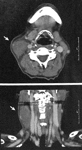
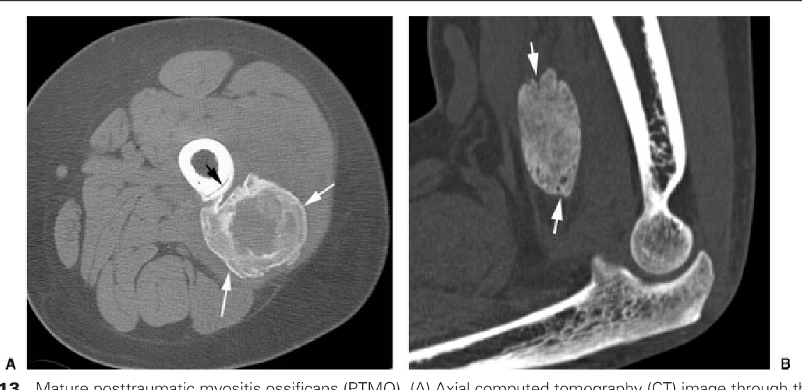
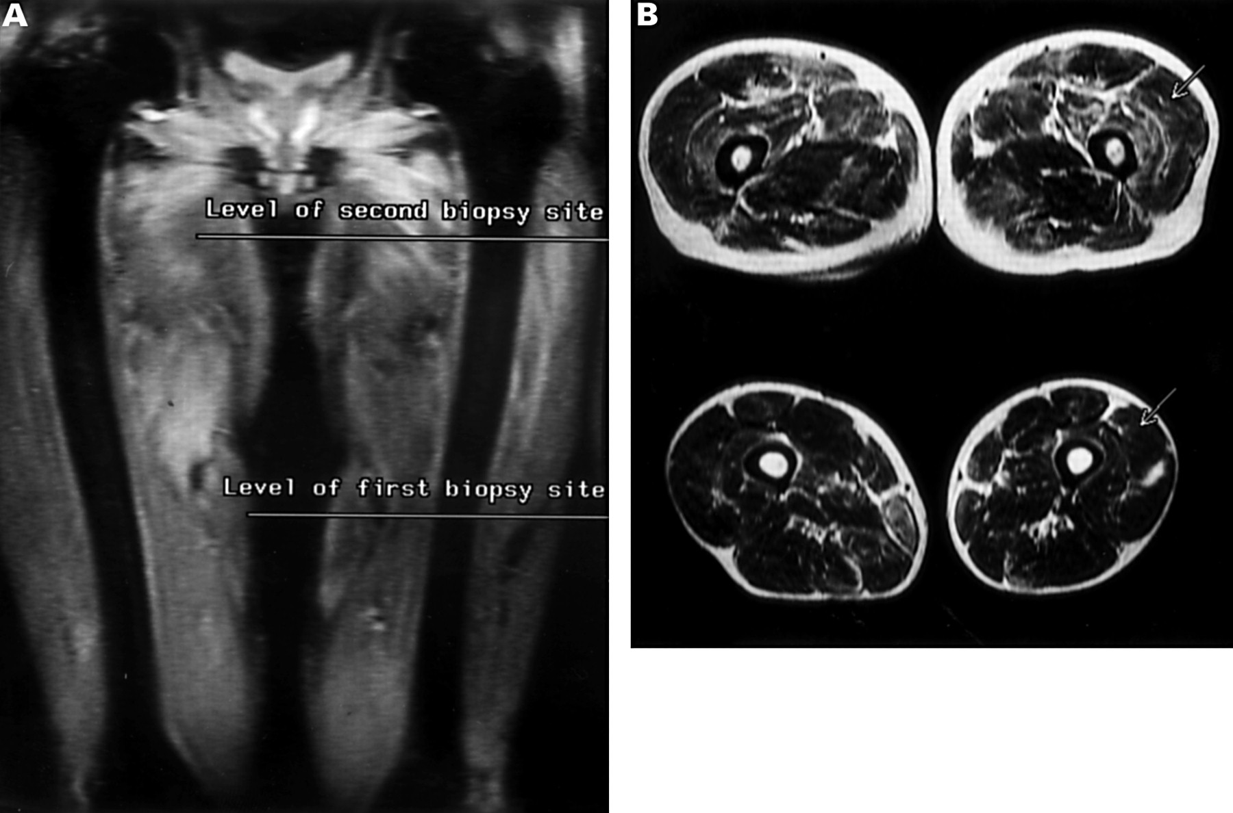
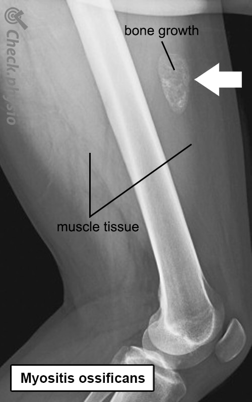
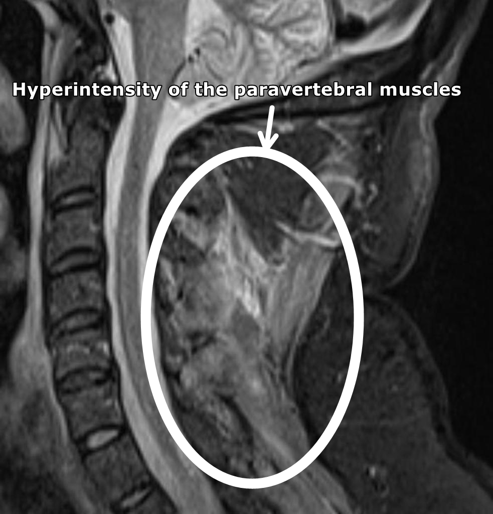
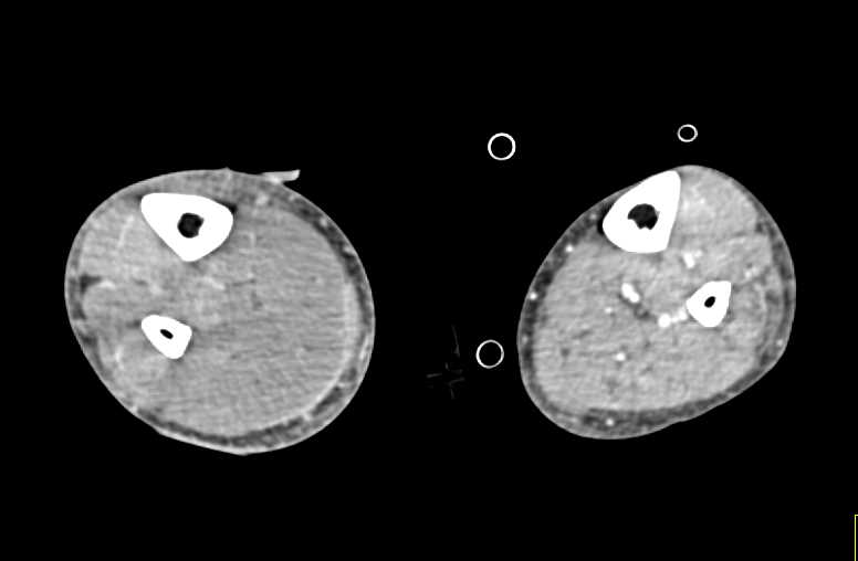
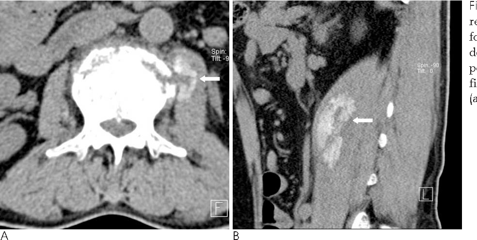
Myositis Pet/ct Radiology


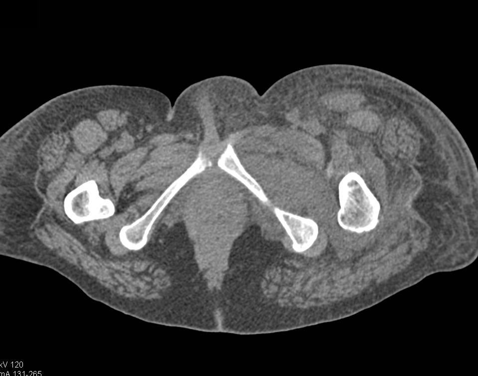


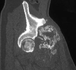


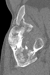
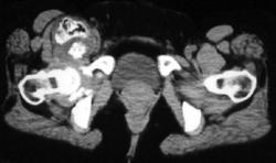
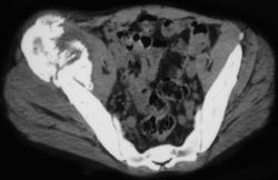
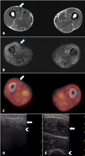
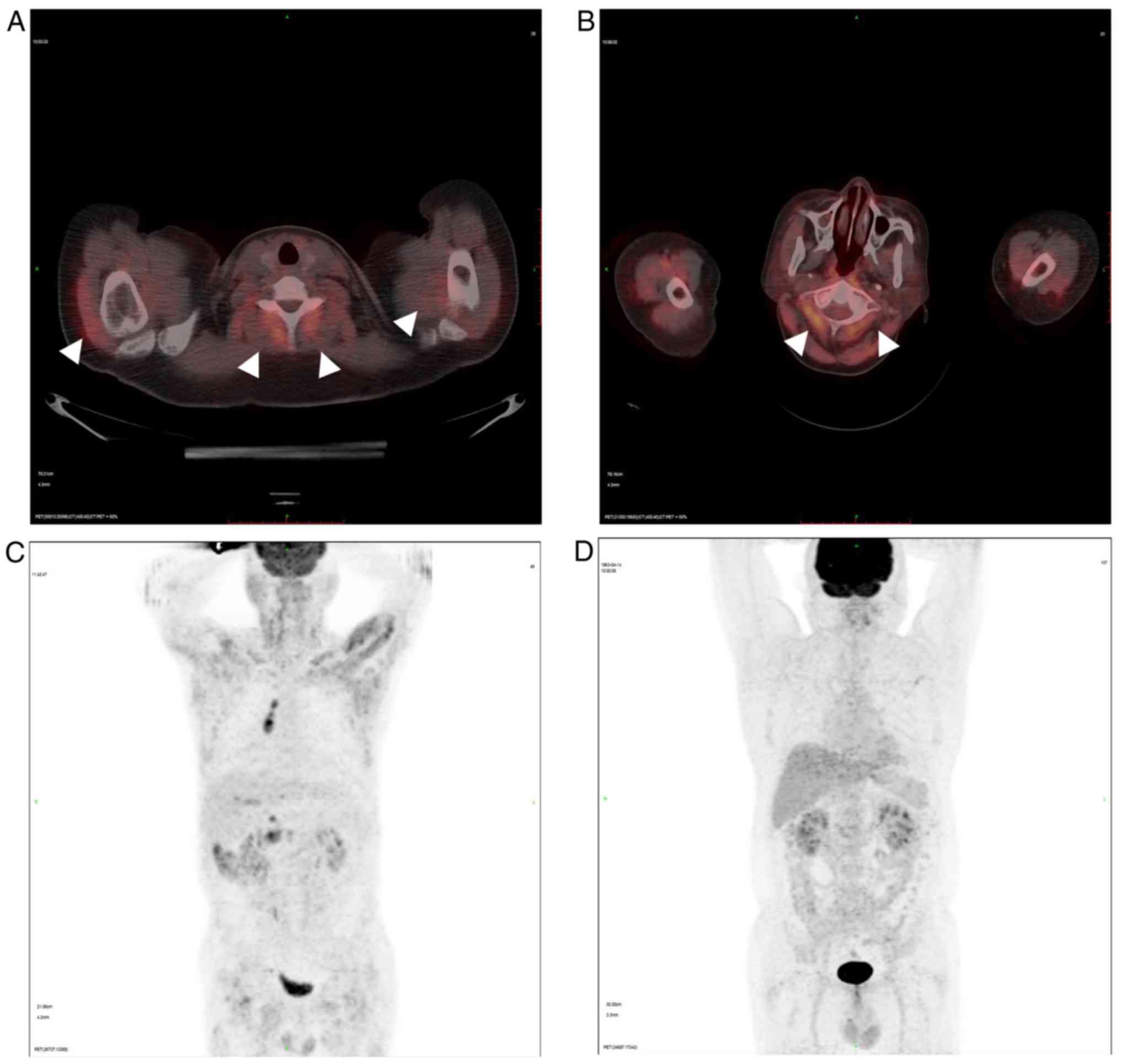
![Signs of skeletal myositis on [⁶⁸Ga]-DOTATOC PET/CT scan. Pathological ...](https://www.researchgate.net/publication/376785786/figure/fig3/AS:11431281214101438@1703386203956/Signs-of-skeletal-myositis-on-Ga-DOTATOC-PET-CT-scan-Pathological-uptake-of-the_Q640.jpg)
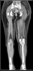
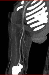
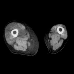
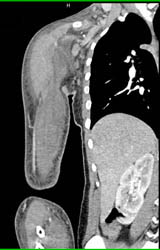
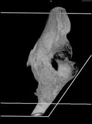
![3D volume rendered PET/CT images of [ 68 Ga]Ga-PVD in murine myositis ...](https://www.researchgate.net/publication/341806419/figure/fig2/AS:899177353576449@1591391995462/3D-volume-rendered-PET-CT-images-of-68-GaGa-PVD-in-murine-myositis-A-and-rat.png)

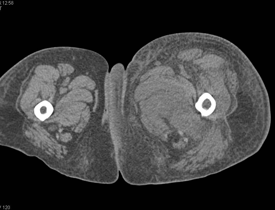
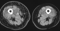
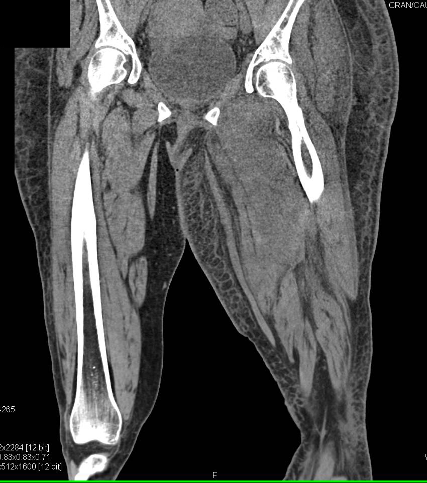


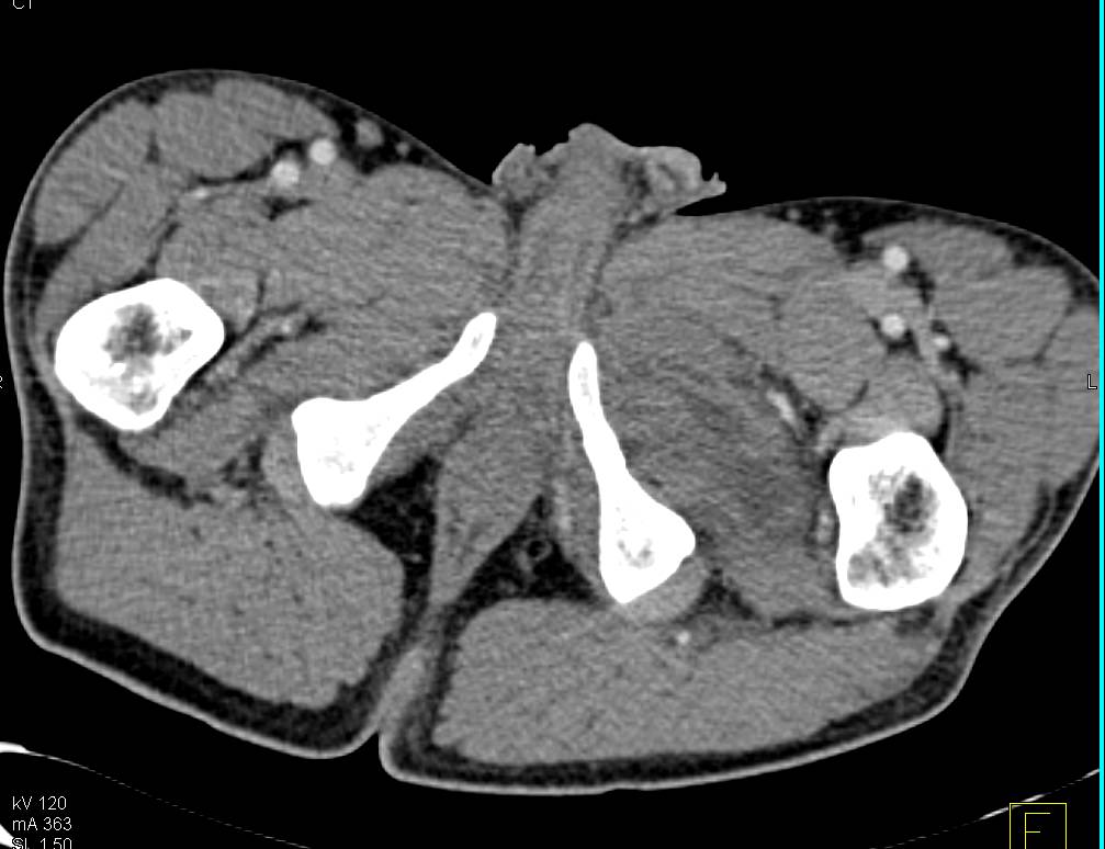
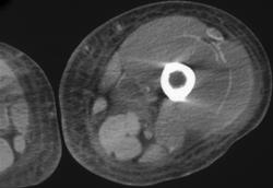
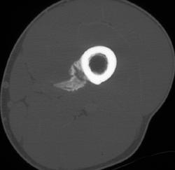
![[18F]FDG-PET/CT in Idiopathic Inflammatory Myopathies: Retrospective ...](https://www.mdpi.com/diagnostics/diagnostics-13-02316/article_deploy/html/images/diagnostics-13-02316-g002.png)


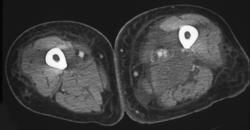
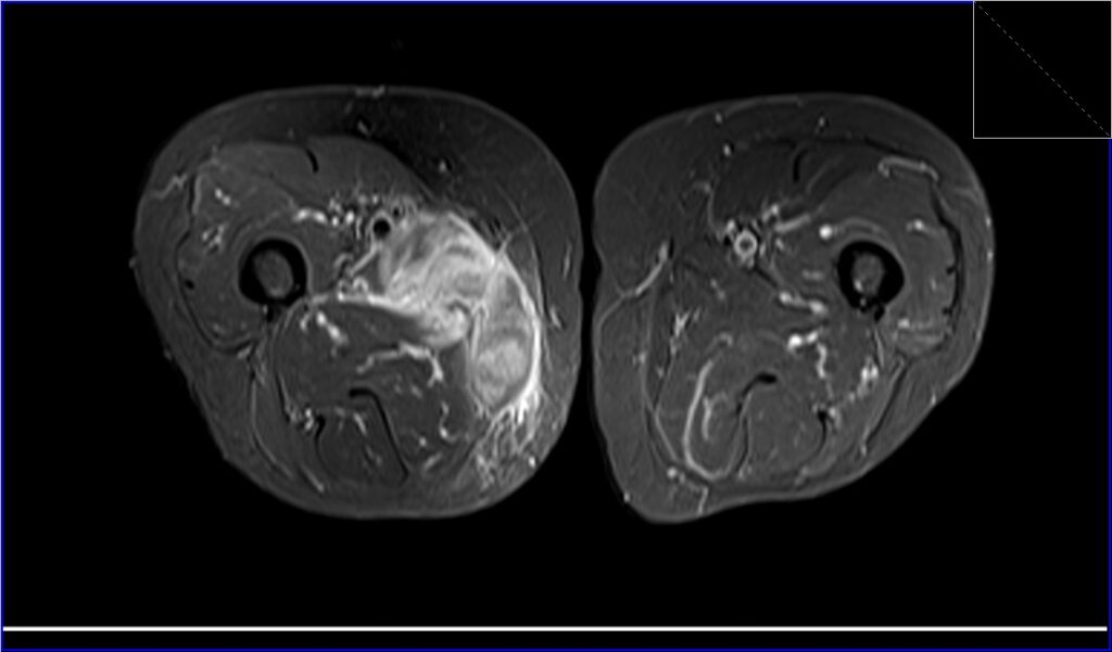
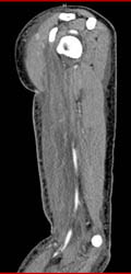
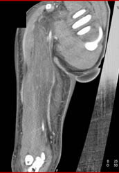
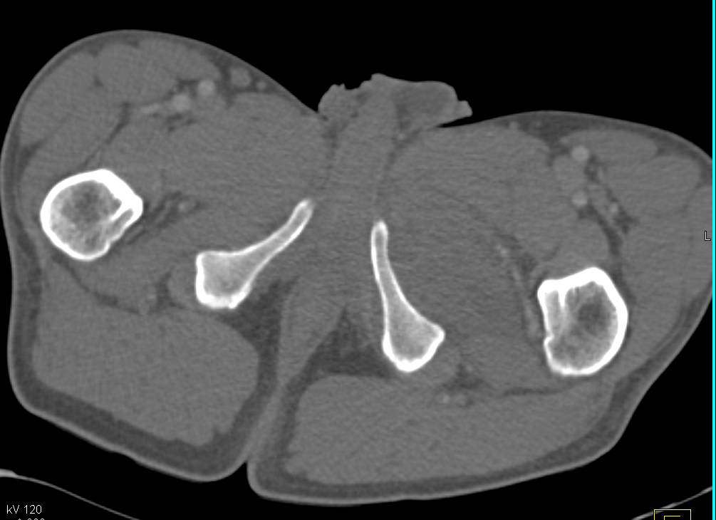
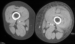
![[18F]FDG-PET/CT in Idiopathic Inflammatory Myopathies: Retrospective ...](https://www.mdpi.com/diagnostics/diagnostics-13-02316/article_deploy/html/images/diagnostics-13-02316-g003.png)


Celebrate the visual poetry of Myositis Pet/ct Radiology through comprehensive galleries of carefully composed images. where technical excellence meets creative vision and artistic expression. creating lasting impressions through powerful and memorable imagery. Discover high-resolution Myositis Pet/ct Radiology images optimized for various applications. Ideal for artistic projects, creative designs, digital art, and innovative visual expressions All Myositis Pet/ct Radiology images are available in high resolution with professional-grade quality, optimized for both digital and print applications, and include comprehensive metadata for easy organization and usage. Our Myositis Pet/ct Radiology collection inspires creativity through unique compositions and artistic perspectives. Professional licensing options accommodate both commercial and educational usage requirements. Time-saving browsing features help users locate ideal Myositis Pet/ct Radiology images quickly. Whether for commercial projects or personal use, our Myositis Pet/ct Radiology collection delivers consistent excellence. Reliable customer support ensures smooth experience throughout the Myositis Pet/ct Radiology selection process. Multiple resolution options ensure optimal performance across different platforms and applications. Cost-effective licensing makes professional Myositis Pet/ct Radiology photography accessible to all budgets. Comprehensive tagging systems facilitate quick discovery of relevant Myositis Pet/ct Radiology content. The Myositis Pet/ct Radiology collection represents years of careful curation and professional standards.