Giant Cell Arteritis Pet/ct


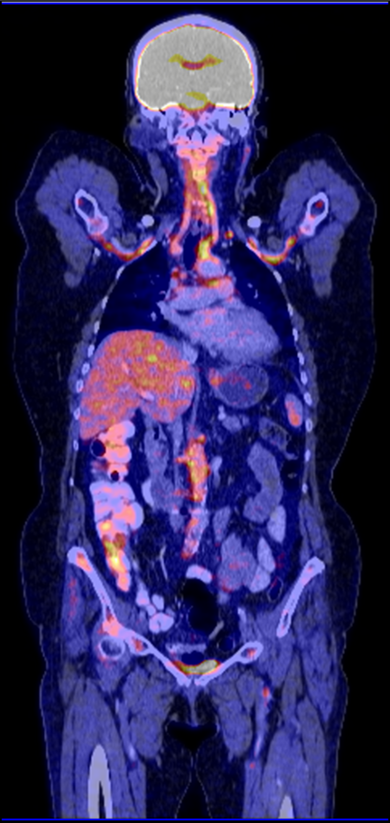
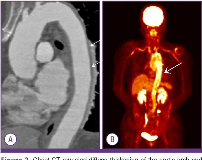
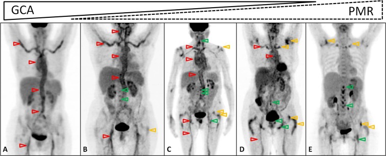
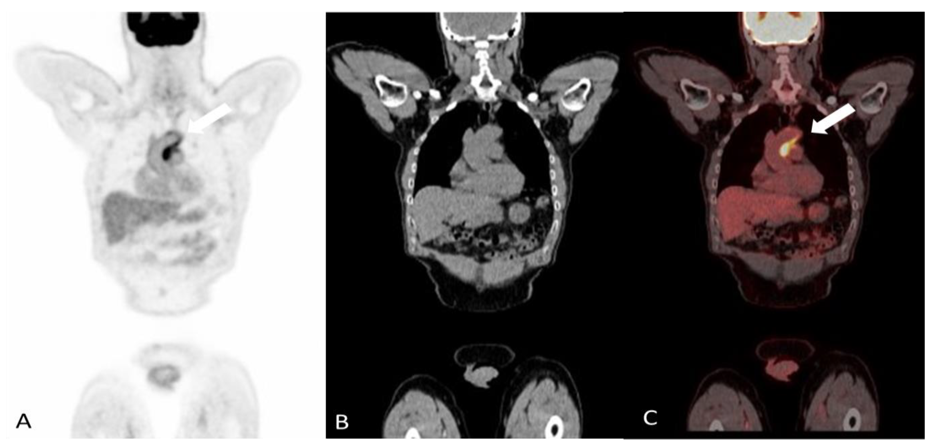
![[18F] FDG-PET/CT in giant cell arteritis (GCA). Panel (A) shows large ...](https://www.researchgate.net/publication/366342020/figure/fig2/AS:11431281115137159@1674774368940/18F-FDG-PET-CT-in-giant-cell-arteritis-GCA-Panel-A-shows-large-vessel-vasculitis_Q320.jpg)


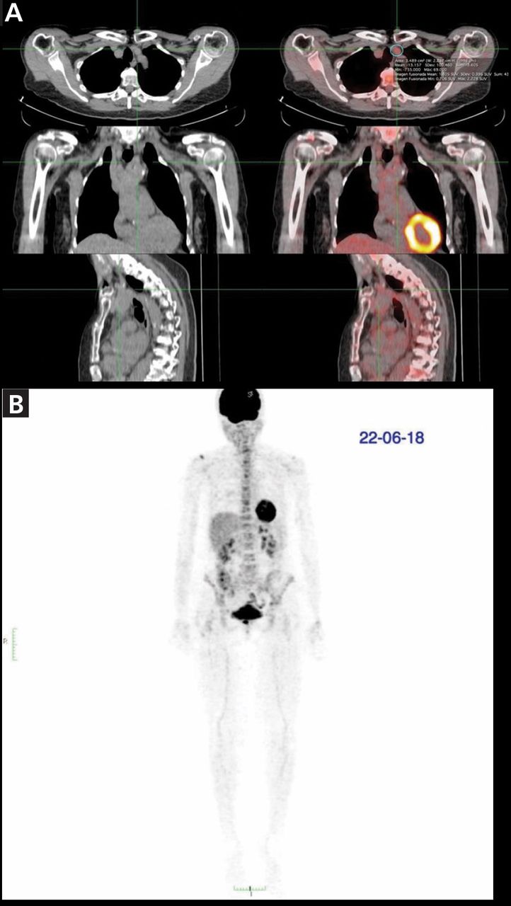
![Giant Cell Arteritis Confirmed by 2-[18F]FDG PET/CT, Missed on Biopsy ...](https://www.aaojournal.org/cms/10.1016/j.ophtha.2023.05.031/asset/71aa849d-aec1-4781-844e-c2d1fcdb8b52/main.assets/gr1_lrg.jpg)


![Frontiers | Case report: Detecting giant cell arteritis in [68Ga]Ga ...](https://www.frontiersin.org/files/Articles/1501790/fimmu-15-1501790-HTML-r1/image_m/fimmu-15-1501790-g003.jpg)


![AB1295 ASSESSING GIANT CELL ARTERITIS ACTIVITY WITH [68GA]GA-DOTA ...](https://ard.bmj.com/content/annrheumdis/83/Suppl_1/1993.2/F1.large.jpg)


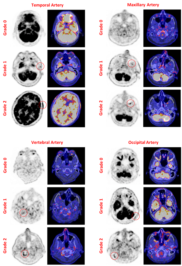
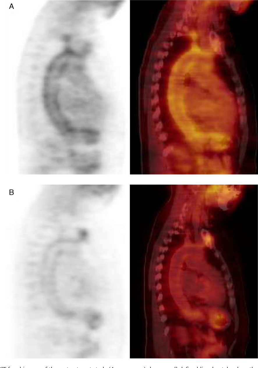
![Visualization of cranial giant cell arteritis with [18F]FDG PET/CT: A ...](https://cdn.ncbi.nlm.nih.gov/pmc/blobs/5a9c/11357830/d9b69919524d/gr3.jpg)
![Visualization of cranial giant cell arteritis with [18F]FDG PET/CT: A ...](https://cdn.ncbi.nlm.nih.gov/pmc/blobs/5a9c/11357830/7c171e5b0f39/gr2.jpg)


![[18F] FDG-PET/CT in giant cell arteritis (GCA). Panel (A) shows large ...](https://www.researchgate.net/publication/366342020/figure/fig1/AS:11431281115154528@1674774368649/Ultrasound-of-the-temporal-artery-with-halo-sign-characterised-by-parietal-thickening_Q320.jpg)
![Visualization of cranial giant cell arteritis with [18F]FDG PET/CT: A ...](https://cdn.ncbi.nlm.nih.gov/pmc/blobs/5a9c/11357830/9e557a5a29d3/gr1.jpg)


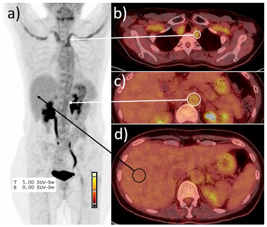


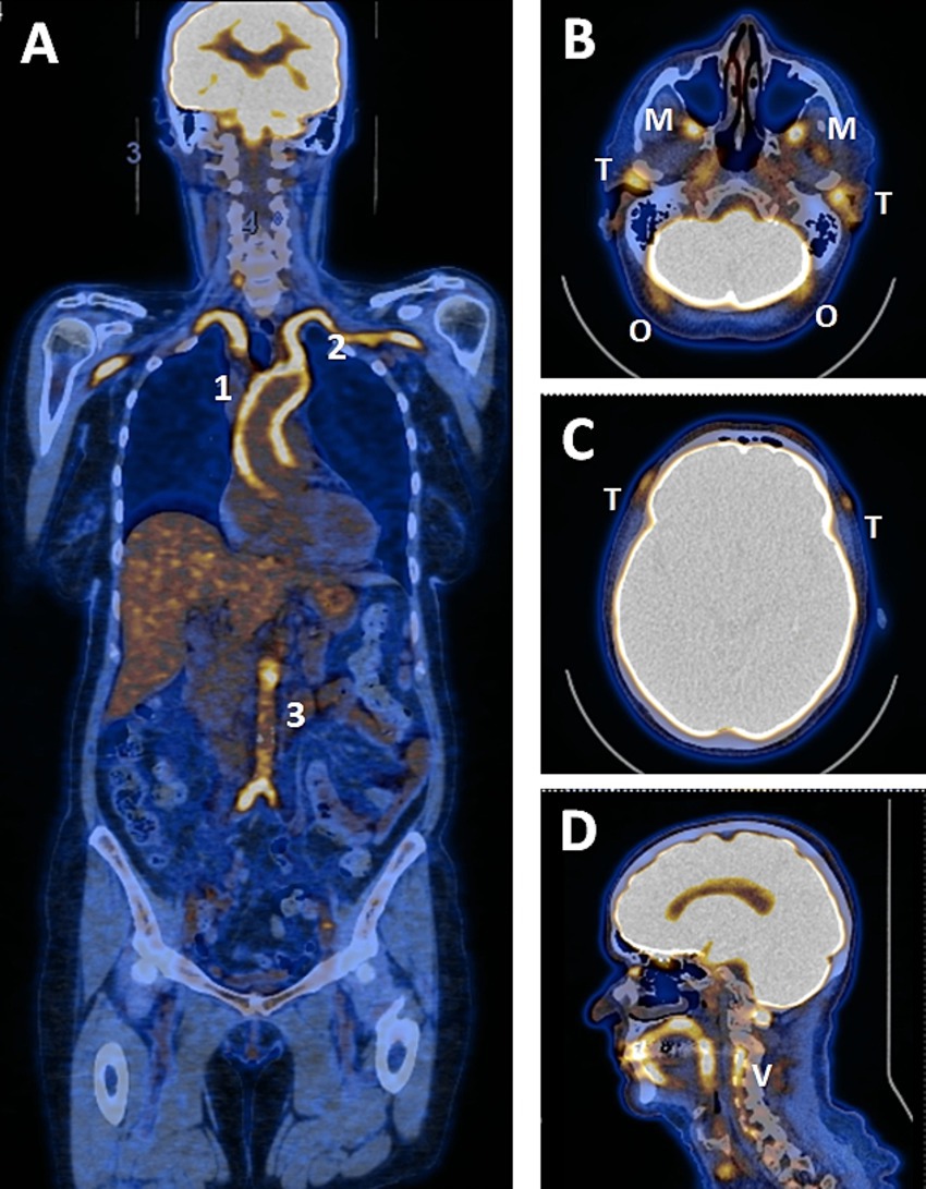
![Coronal [ 18 F]FDG PET (A) and fused PET/CT coronal (B), sagittal (C ...](https://www.researchgate.net/publication/365242540/figure/fig4/AS:11431281096438820@1668151824588/Coronal-18-FFDG-PET-A-and-fused-PET-CT-coronal-B-sagittal-C-and-axial-D.png)


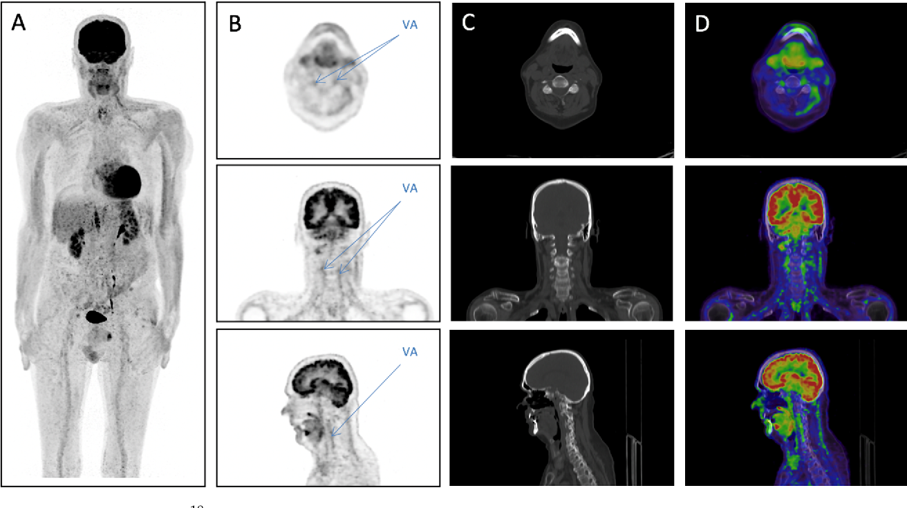
![[ 18 F]FDG PET/CT was performed in a 64 year old patient for a ...](https://www.researchgate.net/publication/356668727/figure/fig1/AS:1098132007976962@1638826481873/18-FFDG-PET-CT-was-performed-in-a-64-year-old-patient-for-a-suspicion-of-giant-cell.png)


![Performance of Deauville Criteria in [18F]FDG-PET/CT Diagnostics of ...](https://pub.mdpi-res.com/diagnostics/diagnostics-13-00157/article_deploy/html/images/diagnostics-13-00157-g001.png?1684460283)


![[68Ga]Ga-DOTA-Siglec-9 PET/CT: A Novel Approach in Assessing Disease ...](https://jnm.snmjournals.org/content/jnumed/65/supplement_2/241691/F2.large.jpg?width=800&height=600&carousel=1)

Document reality with our stunning Giant Cell Arteritis Pet/ct collection of countless authentic images. authentically documenting artistic, creative, and design. designed to preserve authentic moments and stories. Discover high-resolution Giant Cell Arteritis Pet/ct images optimized for various applications. Suitable for various applications including web design, social media, personal projects, and digital content creation All Giant Cell Arteritis Pet/ct images are available in high resolution with professional-grade quality, optimized for both digital and print applications, and include comprehensive metadata for easy organization and usage. Our Giant Cell Arteritis Pet/ct gallery offers diverse visual resources to bring your ideas to life. Our Giant Cell Arteritis Pet/ct database continuously expands with fresh, relevant content from skilled photographers. Regular updates keep the Giant Cell Arteritis Pet/ct collection current with contemporary trends and styles. Instant download capabilities enable immediate access to chosen Giant Cell Arteritis Pet/ct images. Advanced search capabilities make finding the perfect Giant Cell Arteritis Pet/ct image effortless and efficient. Cost-effective licensing makes professional Giant Cell Arteritis Pet/ct photography accessible to all budgets. Multiple resolution options ensure optimal performance across different platforms and applications. Time-saving browsing features help users locate ideal Giant Cell Arteritis Pet/ct images quickly.