Please enter url.
Login
Logout
Please enter url.
Loading ...
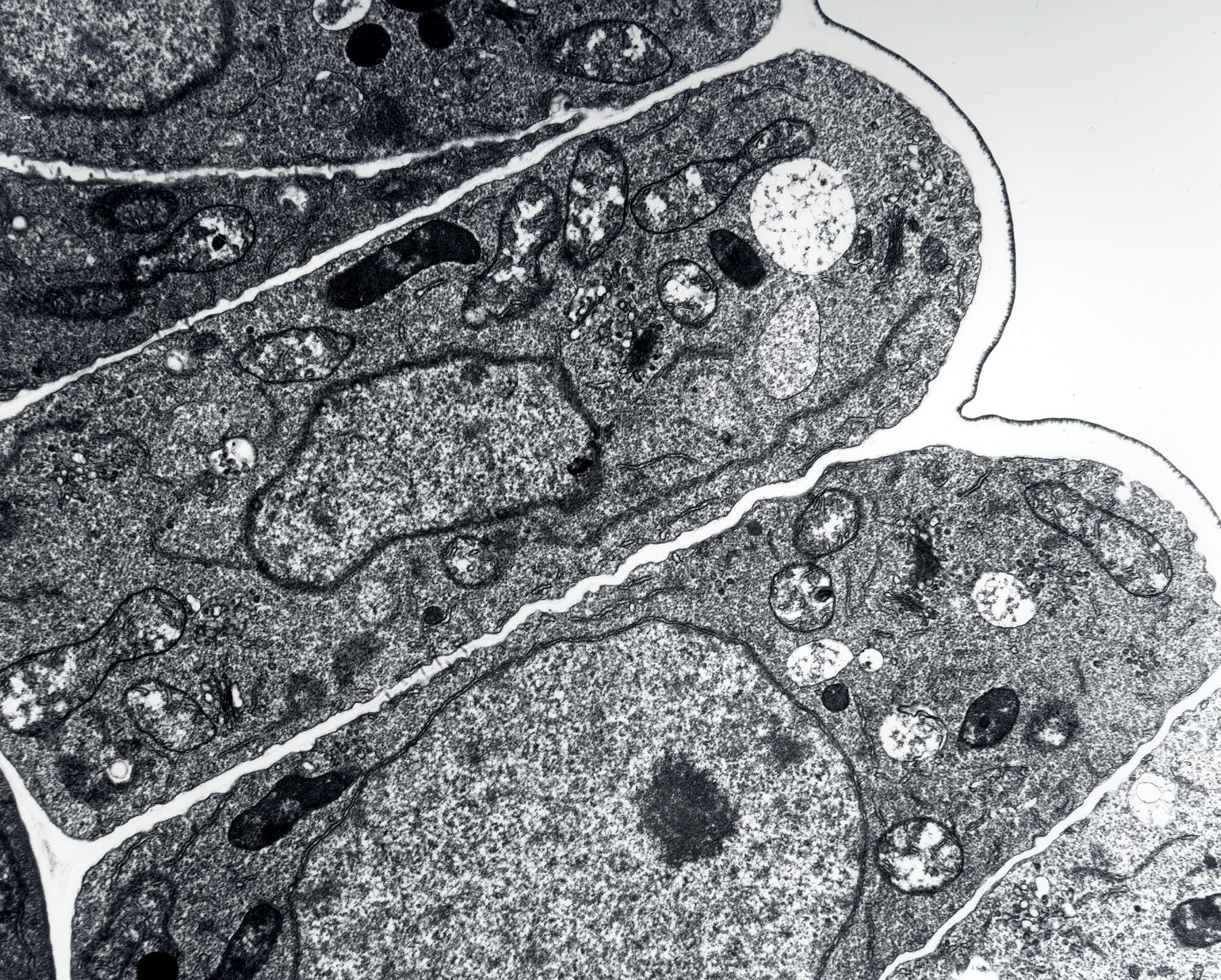
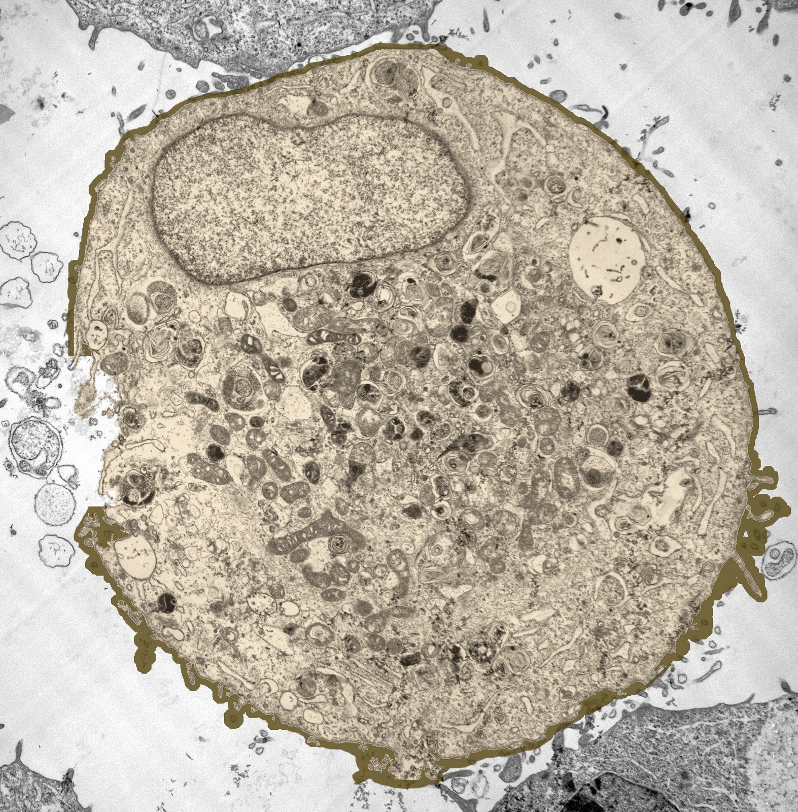
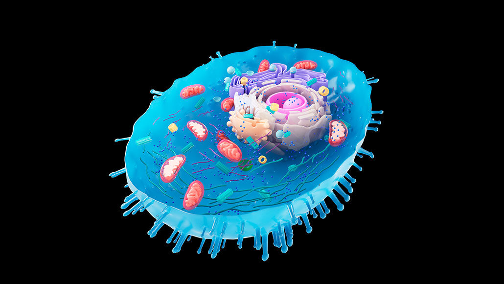
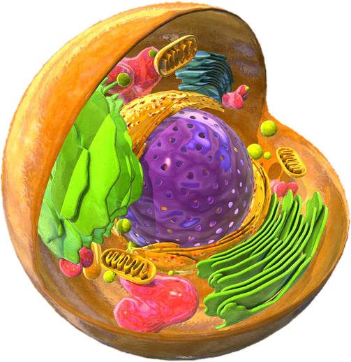
Electron Micrograph Of Eukaryotic Cell
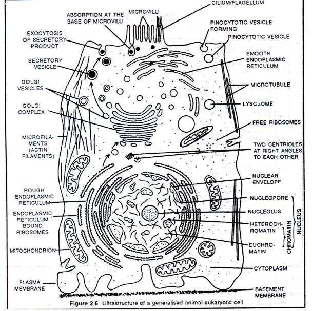
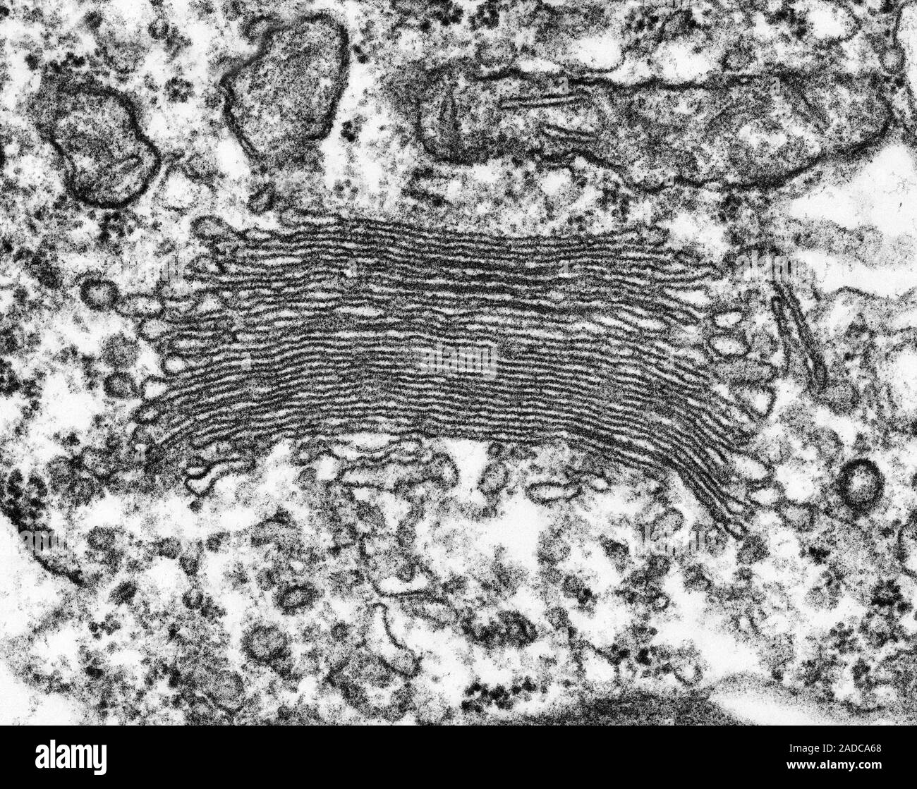
Electron Micrograph Diagram
Diagram Of An Electron Microscope
Electron Micrograph Diagram
Vacuole Electron Micrograph Animal Cell
Plant Cell With Microscope at Franklin Moffet blog
The Cell Form 1 Biology Notes Simple Diagram Of Golgi Apparatus
Ribosomes Animal Cell Model
Electron Microscope Images Of Cells
Cell - Upper sec Science
Cell Wall And Cell Membrane Microscope
Cells In A Microscope
Golgi Apparatus Microscope
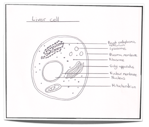
Hepatocyte (liver cell)| various organelles including the nucleus ...
Eukaryotic Micrograph Images Diagram | Quizlet
Plant Cell With Microscope at Franklin Moffet blog
[Figure, Transmission Electron Micrograph of Rough Endoplasmic ...
How Does A Plant Cell Look Like Under The Microscope at Elaine Ortiz blog
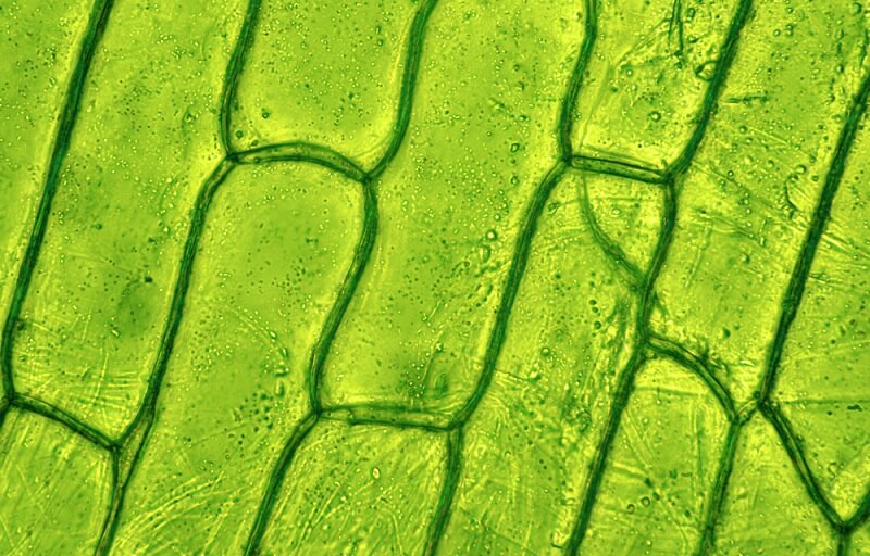
Eukaryotic Plant Cell Under Microscope
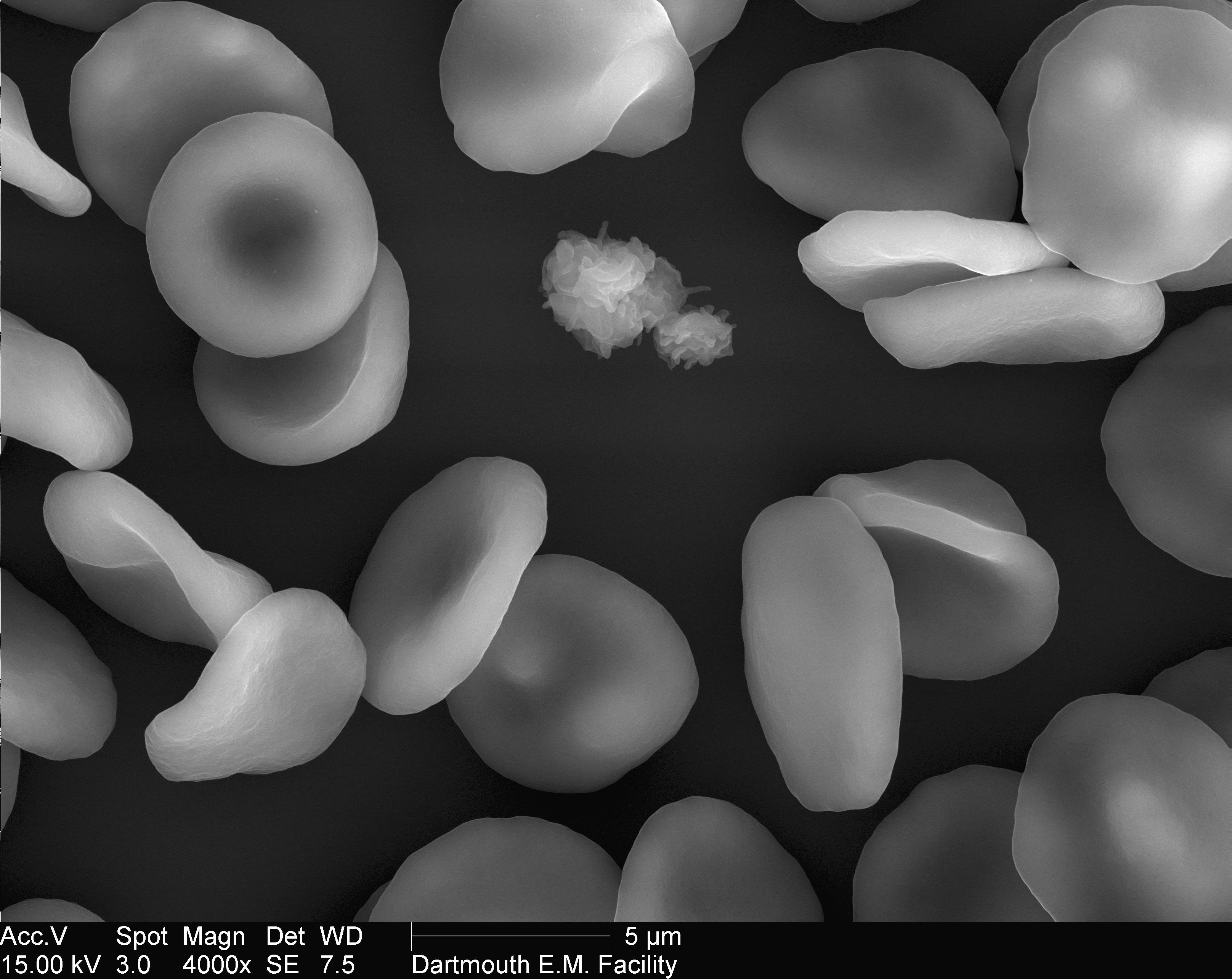
Scanning Electron Microscope Cells
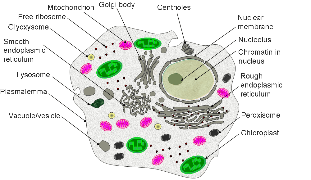
Eukaryotic Cell Diagram Labeled
Electron microscope, Scanning electron microscope, Microscopy
[DIAGRAM] Typical Eukaryotic Cell Diagram - MYDIAGRAM.ONLINE
Labeled Animal Cell Under Microscope
Bacterial Cells Under A Microscope
Simple Eukaryotic Cell Diagram
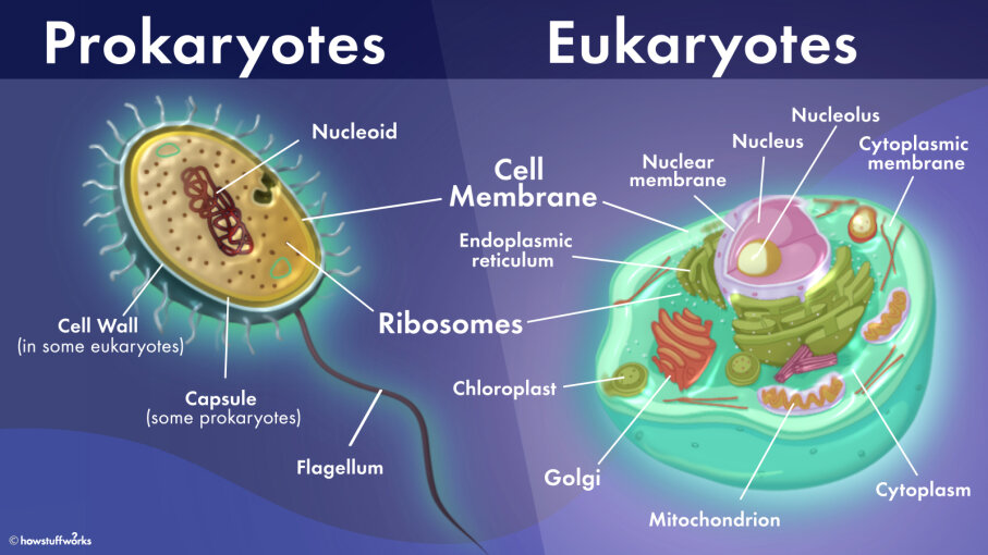
Eukaryotes
Lysosome Microscope
Eukaryotic Cell Parts Diagram
Skills: Cell Origin & Ultrastructure (1.2.9) | DP IB Biology: SL ...
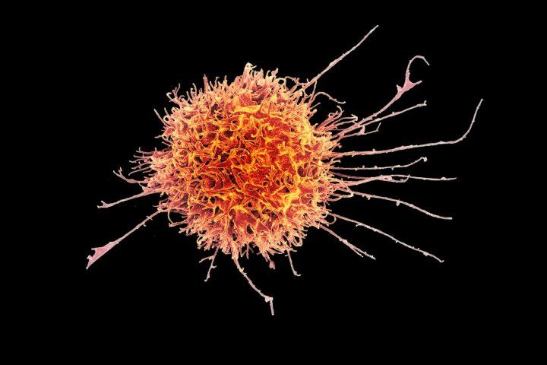
Leukemia blood cells under a Color scanning electron micrograph. Red ...
Eukaryotic Animal Cell Simple Diagram
BIO120Concepts of BiologyUnit 2 Lecture Part One Cel.docx
Eukaryotic Cell Diagram Unlabeled
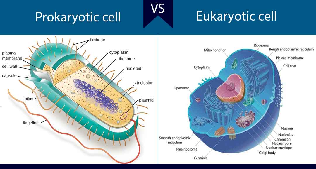
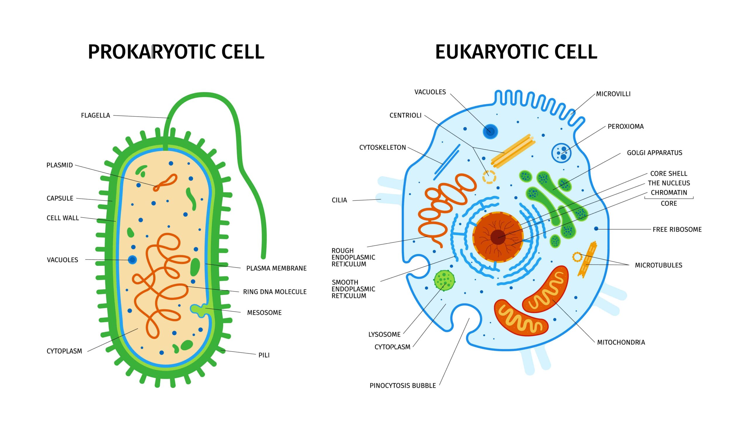
Prokaryotic And Eukaryotic Cells Lesson
Albums 104+ Images Picture Of Cytoplasm In A Plant Cell Latest
diklatkerja | Sejarah Perkembangan Pandangan pada Biologi, Ilmu Tentang ...
Prokaryotic Vs Eukaryotic Cells Worksheet Key
Organelles In Eukaryotic Cells Worksheet
Bio 1.1.2 - NC Bio
Animal Cell Under Microscope Diagram - Get More Anythink's
Electron Micrograph Diagram
Video: Correlative Light and Electron Microscopy CLEM as a Tool to ...
CIL:7600, Halteria grandinella, cell by organism, eukaryotic cell ...
chapter 7 - cell structure and function | Quizlet
CIL:7600, Halteria grandinella, cell by organism, eukaryotic cell ...
chapter 7 - cell structure and function | Quizlet
chapter 7 - cell structure and function | Quizlet
This 'gastric rainbow', is a microscopic section through a mouse ...
2,337 Mitochondria Picture Stock Photos, High-Res Pictures, and Images ...
This 'gastric rainbow', is a microscopic section through a mouse ...
New £125M world-first diffraction and imaging electron microscope to ...
B1 - Cell Level Systems Flashcards | Quizlet
Bio Ch 21 Flashcards | Quizlet
Biology 114 chapter 4 extra questions Flashcards | Quizlet
biology paper 1 Flashcards | Quizlet
Bio exam 2 chp 22 Flashcards | Quizlet
Nucleosome conformational variability in situ and in vitro
Chapter 2 - microbio Flashcards | Quizlet
Macrophage, SEM - Stock Image - C057/8601 - Science Photo Library ...
Microtubules. Transmission electron micrograph of purified … [158264961 ...
Macrophage, SEM - Stock Image - C057/8601 - Science Photo Library ...
Paper 1 | Flashcards
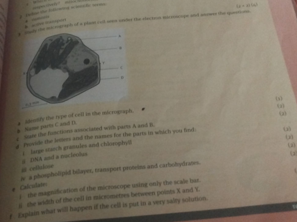
3 Study the micrograph of a plant cell seen | StudyX
3.1 Study the diagram of a cell seen under | StudyX
Stem Cell Medical Background Royalty-Free Images, Stock Photos ...
Video: Micropatterning Transmission Electron Microscopy Grids to Direct ...
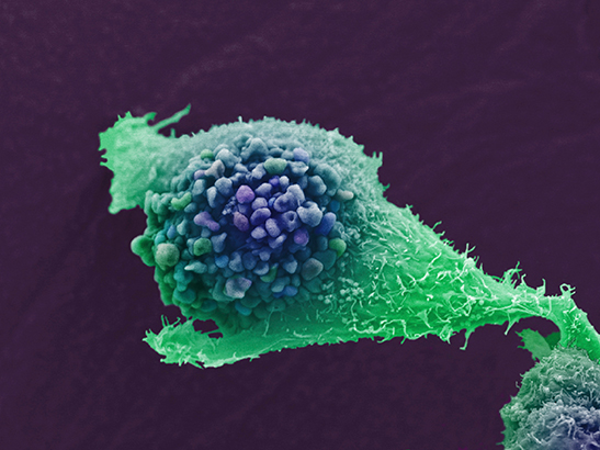
AACR 2022: New 'killer' immunotherapy shows early promise in range of ...
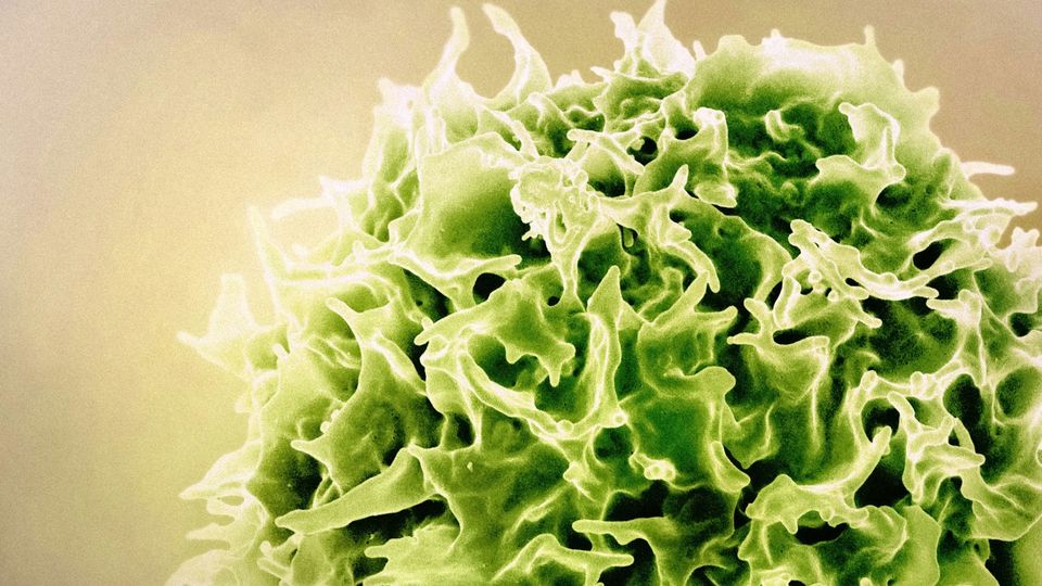
New combination therapy exploits ‘natural killer’ cells to destroy head ...
Video: Preparation of Graphene-Supported Microwell Liquid Cells for In ...
A drawing from a micrograph of a cell | StudyX
Why this is the worst flu season in 15 years
White Blood Cells Use Force To Dislodge Bacteria | Technology Networks
ICR responds to NICE decision not to recommend olaparib for advanced ...
High voltage electron microscopy of cytoskeletal structures in whole ...
Table 1 - Reprogramming of Stem Cell Activity to Convert
Dr. David Meierhofer
CIL:19538, Didinium nasutum, cell by organism, eukaryotic cell ...
Video: Preparation of Prokaryotic and Eukaryotic Organisms Using ...
Figure 2 from Correlative super-resolution fluorescence and electron ...
Structural mechanism of LINE-1 target-primed reverse transcription
Visualization of Plant Cell Wall Epitopes Using Immunogold Labeling for ...
CIL:39250, Didinium nasutum, Paramecium sp., cell by organism ...
Exploring the Growth and Dynamics of Electron Microscopy - Investors ...
How HIV research has reshaped modern medicine — Harvard Gazette
Buy Lokalisierung von Nanopartikeln in Zellen mittels ...
Fauna of India Checklist: Phylum Chaetognatha | Request PDF
Venn Diagramcomparing Prokaryotesand Eukaryotes Goeswith Cells Part 1-1 ...
Video: Analysis of Minerals Produced by hFOB 1.19 and Saos-2 Cells ...
Fungi of relevance to dentistry - Essential Microbiology for Dentistry ...
CIL:10804, endocrine cell. CIL. Dataset
Protein Folding Flashcards | Quizlet
Visualization of Plant Cell Wall Epitopes Using Immunogold Labeling for ...
İnsan Kemiği | Microscopic photography, Scanning electron microscope ...
Visualization of Plant Cell Wall Epitopes Using Immunogold Labeling for ...
4 children surpass a year of HIV remission after treatment pause: Study ...
Cell division | EBSCO Research Starters
Cryo‐Immunogold Electron Microscopy - Peters - 2006 - Current Protocols ...
Study of Oxidative Stress and Apoptotic Genes in SW480 Cell Lines ...
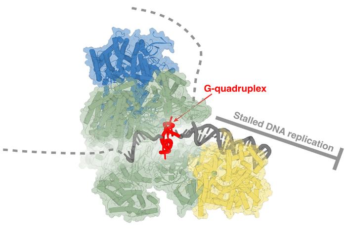
Revolutionary Cryo-Electron Microscopy Unlocks Secrets of DNA Replication
Celula animal, partes y composición | Animal cell, Human cell structure ...
Database of articles cited by 3DEM data entries - EMN Papers
Cell Organelles | HHMI's Beautiful Biology
Figure 1: Transmission electron microscope | StudyX
Cryo-electron microscopy reveals hidden mechanics of DNA replication ...
Unit 4: Cell Structure and Function – TRU Human Anatomy & Physiology I
The Structural Framework In A Cell Is The: | insightx
Scientist Looking Microscope With Dark Background Royalty-Free Images ...
The Structure and Function of Cells Flashcards | Quizlet
6 Bright Microscopic Images Of Life | Microscopic photography ...
Liquid Phase Electron Microscopy and Spectroscopy of Biological ...
CIL:40317, Homo sapiens, epithelial cell, cervical carcinoma. CIL. Dataset
Lab 6 – Use of the microscope Flashcards | Quizlet
Lassa fever | EBSCO Research Starters
From music to molecules: Fred Hutch’s Dr. Melody Campbell’s lab is one ...
Table 1 - Reprogramming of Stem Cell Activity to Convert
Wolbachia | EBSCO Research Starters
Characterization and Growth of Polymorphic Rickettsia felis in a Tick ...
Electron Microscope Eukaryotic Cell
Cell Membrane Electron Micrograph
Liver Cell Electron Micrograph
Electron Micrograph of an Animal Cell
Eukaryotic Cell Under Electron Microscope
Electron Micrographs of Cell Organelles
Plant Cell Micrograph
Electron Micrograph of Plant Cell Labeled
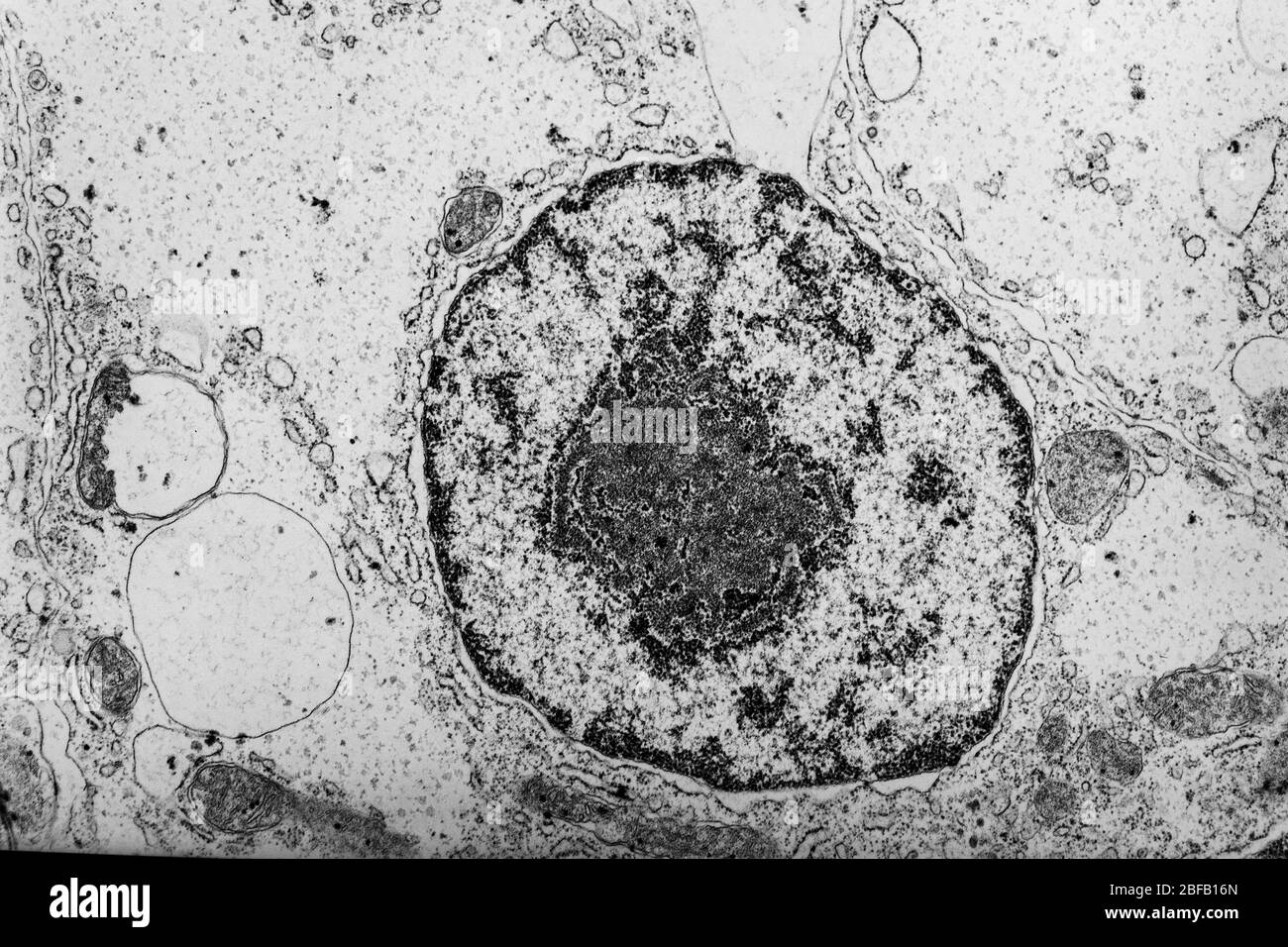
Cell Nucleus Electron Micrograph
Plasma Cell Electron Micrograph
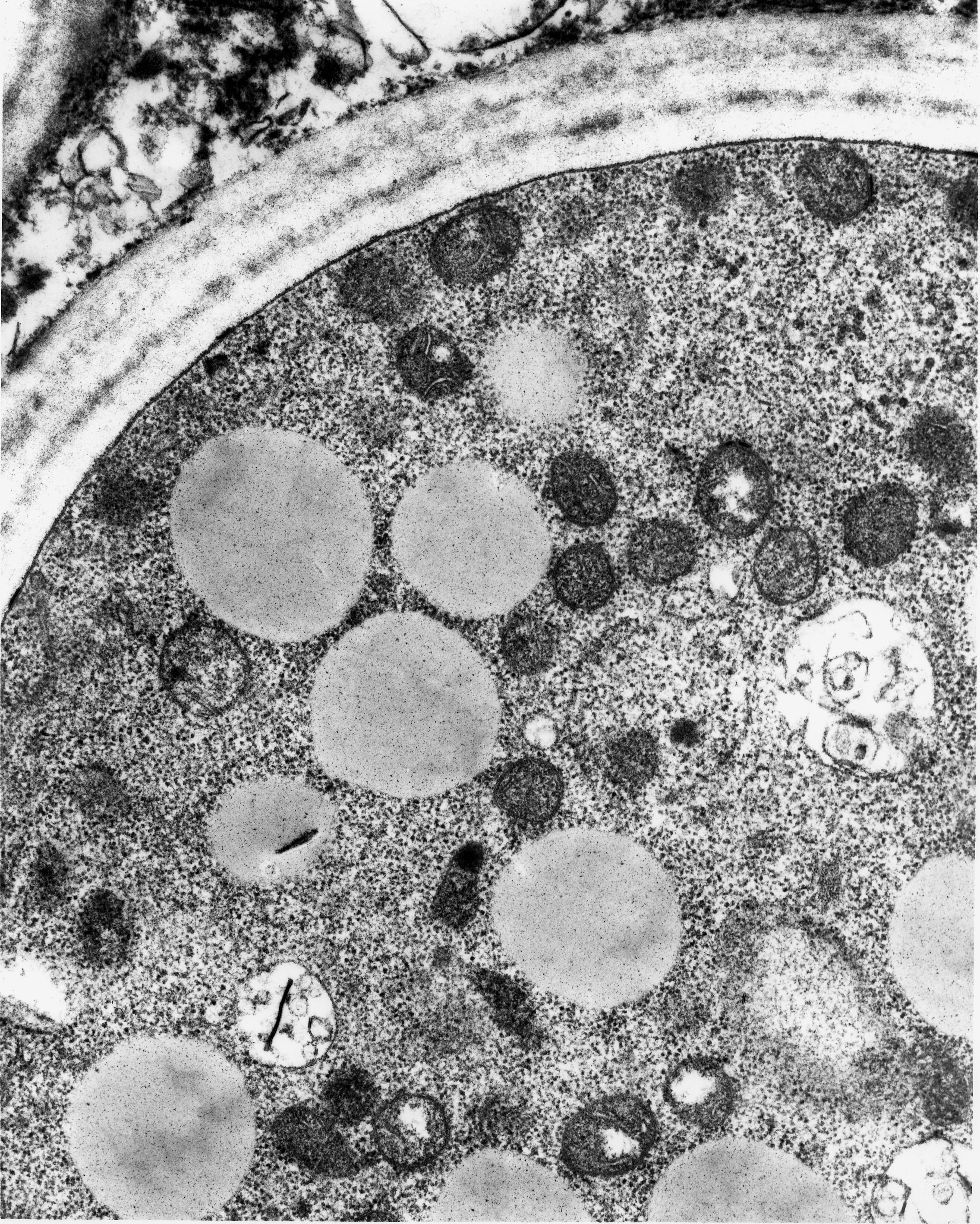
Cell Wall Electron Micrograph
Lysosome Microscope
Plant Cell Diagram Microscope
Prokaryotic Cell Electron Micrograph
Transmission Electron Micrograph
Labelled Eukaryotic Cell Electron Micrograph
Eukaryotic Cell Diagram Unlabeled
Ribosomes Under Electron Microscope
Eukaryotic Cell Structure Drawing
Tissue Cells Under Microscope
Human Cell Nucleus
Mitosis Electron Microscope
Eukaryotic Cell Ultrastructure
Label Your Own Diagram and Electron Micrograph of a Eukaryotic Cell
Eukaryotic Cell Electron Micrograph for an Animal Cell
Sem Image of Mitochondria
Eukaryote Under Microscope
Centriole Electron Micrograph
Electron Micrographs of Eukaryotic Cells E
Vacuole Electron Micrograph
Prokaryote Microscope
Chloroplast Electron Micrograph
Typical Eukaryotic Cell
Golgi Apparatus Electron Micrograph
Eukaryotic Celll Electron Micrograph Annotated
Peroxisome Cell Structure
Animal Cell through Microscope
Eukaryotic Cells From Diagrams and Electron Micrographs
Animal Cell Electron Micograph
Eukaryotic Cell DNA Electron Micrograph
Micrograph of the Cell and Its Sections
Animal Cell Microscope
Centrioles Electron Microscope
Mitochondria électron Micrograph
Electron Micrograph of Golgi Apparatus
Electron Micrograph of Bacteria
Eukaryotic Cell Drawing Simple
Chromosomes Electron Micrograph
Eukaryotic Animal Cell Model
Eukaryotic Cell Characteristics


![[DIAGRAM] Typical Eukaryotic Cell Diagram - MYDIAGRAM.ONLINE](https://www2.mrc-lmb.cam.ac.uk/microscopes4schools/media/cells_proeuk.jpg)


_945x532.jpg)