Cns Lymphoma Pet/ct

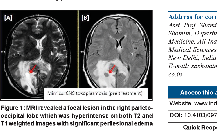
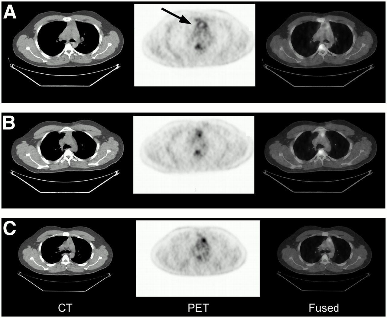
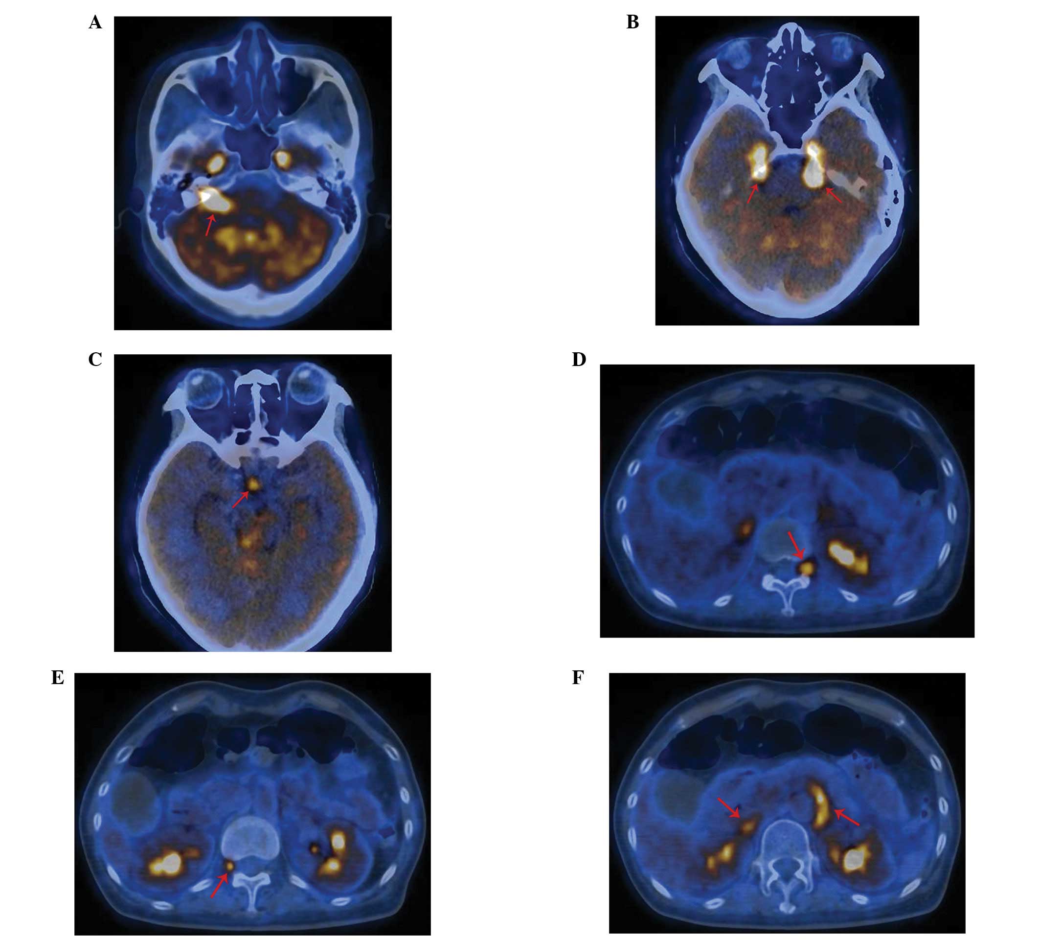
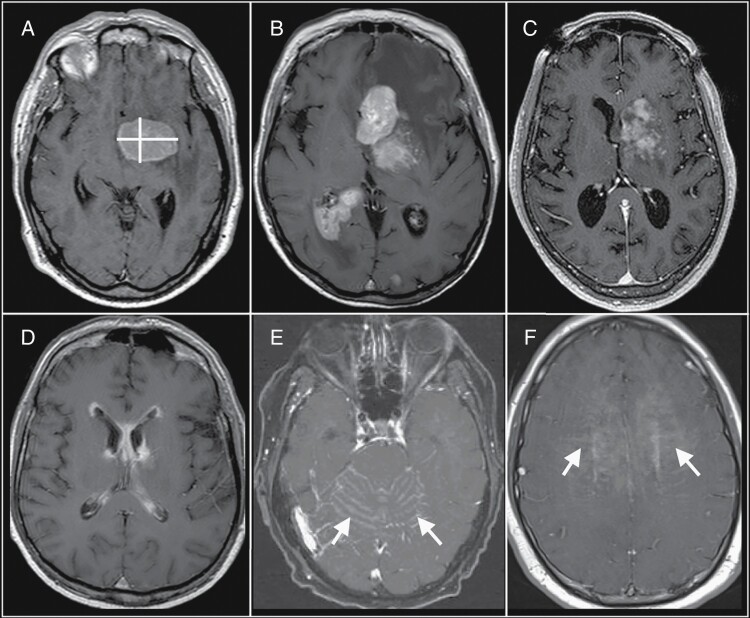
![[18F]Fluoromethylcholine PET/CT for CNS lymphoma... | F1000Research](https://f1000researchdata.s3.amazonaws.com/manuscripts/76872/e790ee36-19e6-45c9-8f95-2aa543eead7c_figure1.gif)


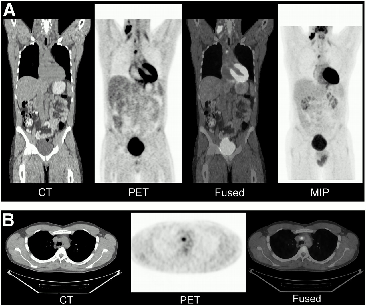
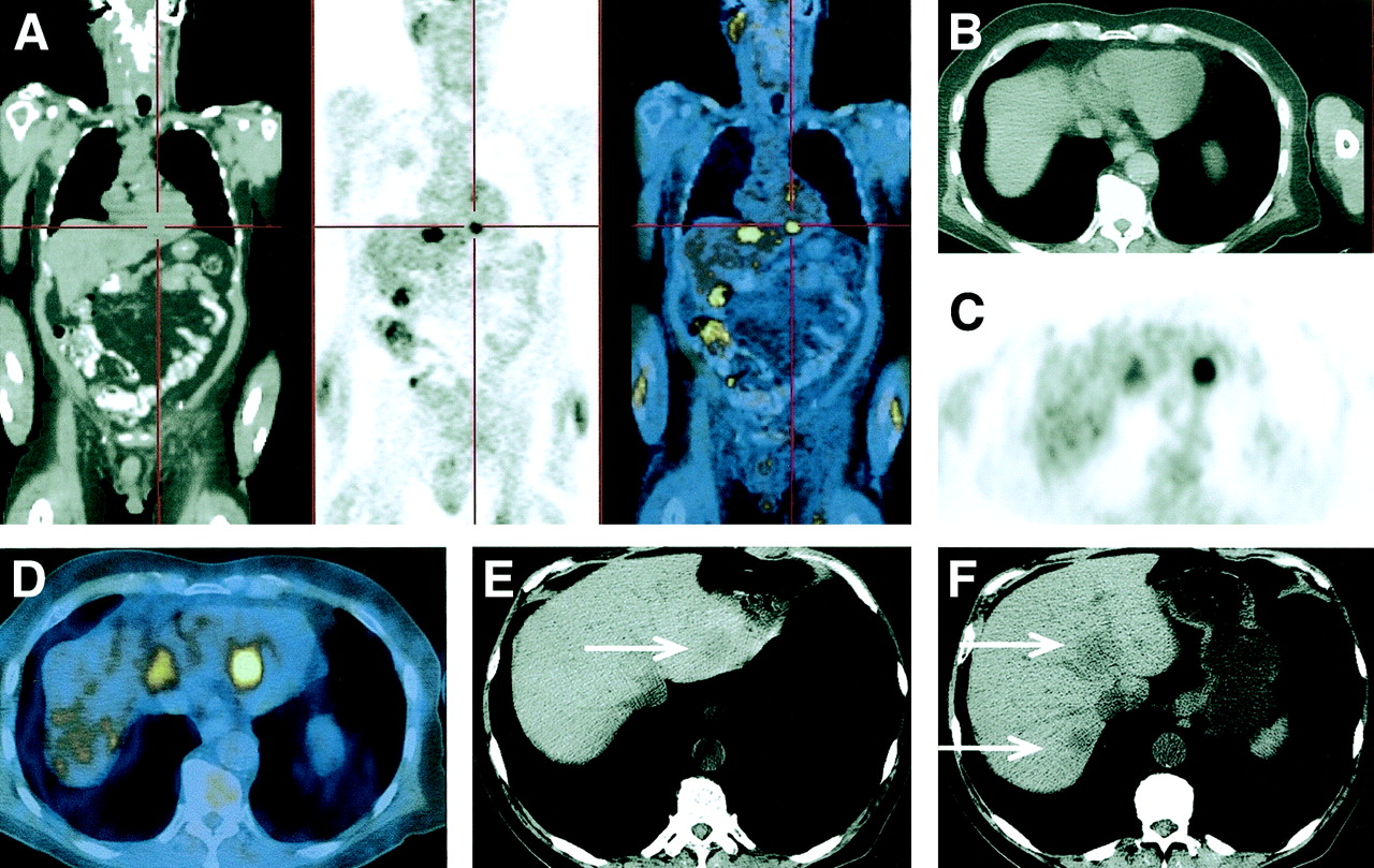
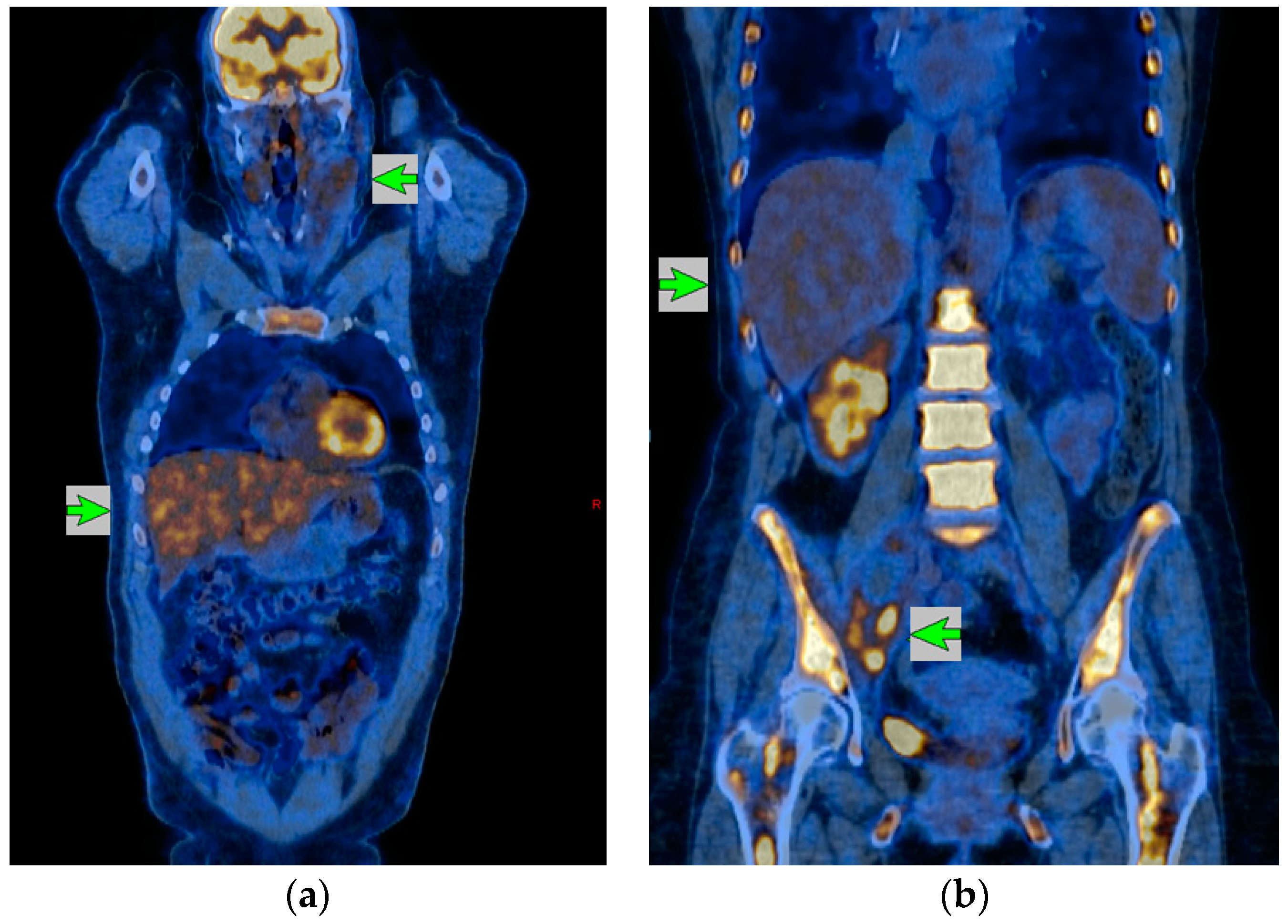
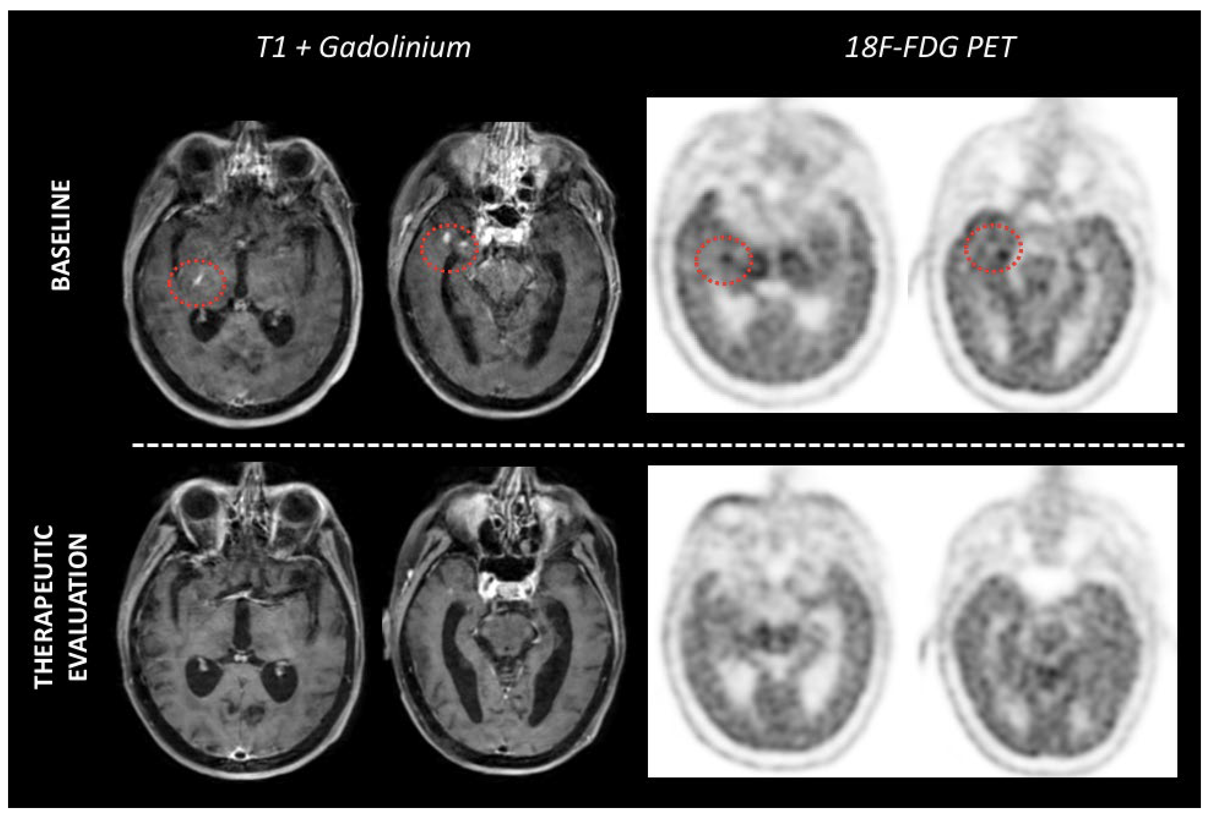
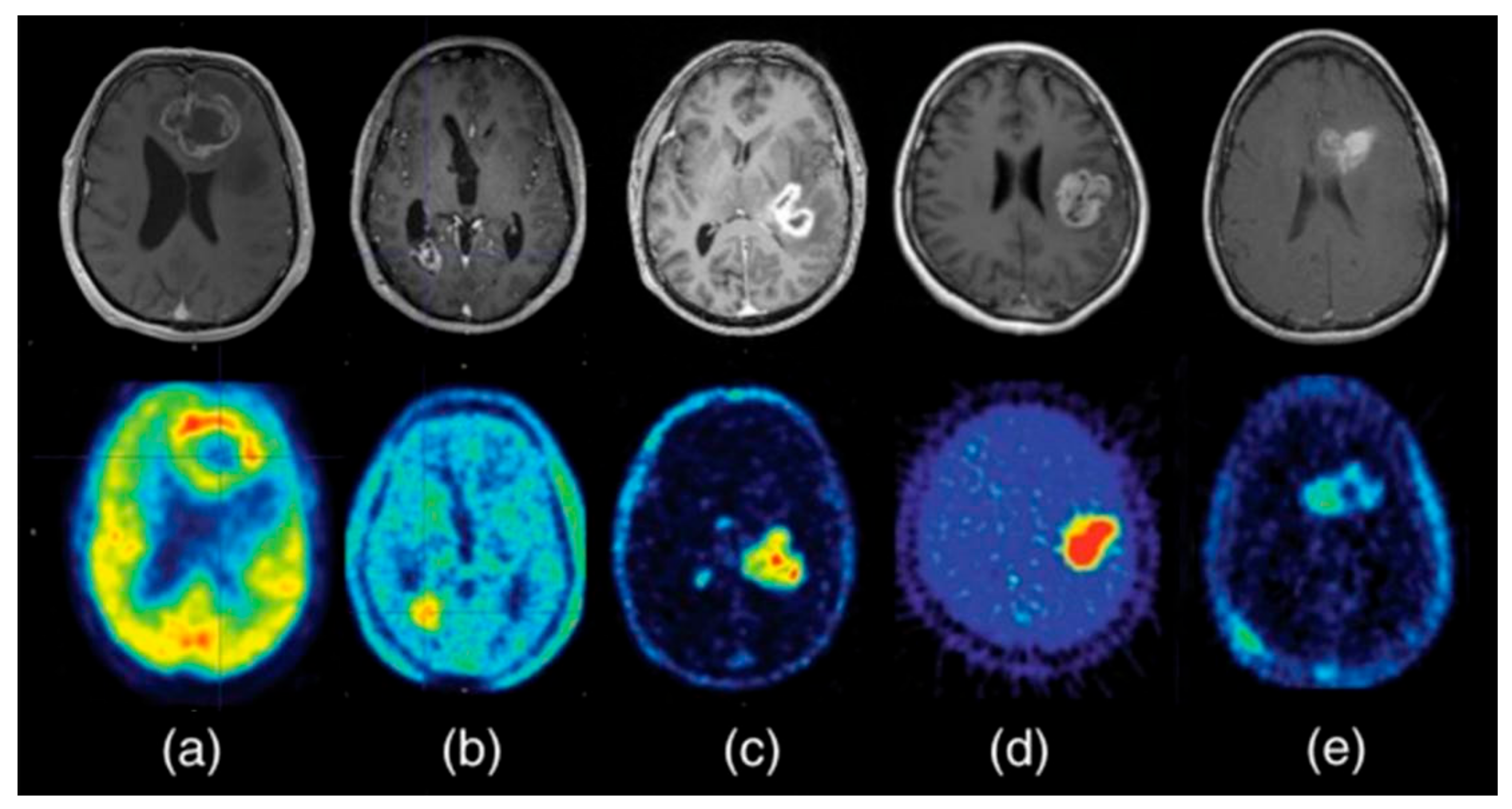
![[18F]FDG PET/CT in suspected primary CNS lymphoma: Systemic involvement ...](https://static.elsevier.es/multimedia/22538089/unassign/S2253808925000692/v1_202506120422/en/main.assets/gr2.jpeg?idApp=UINPBA00004N)


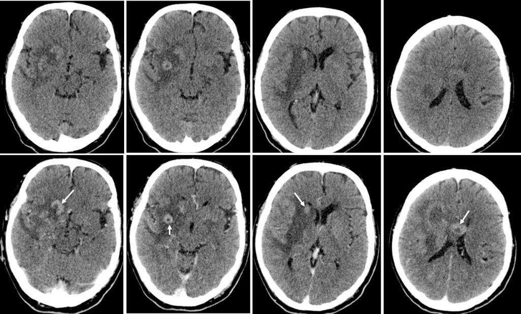


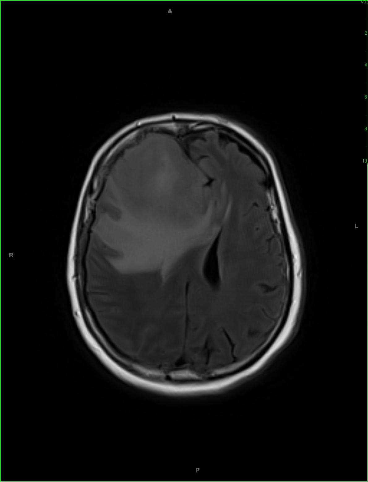
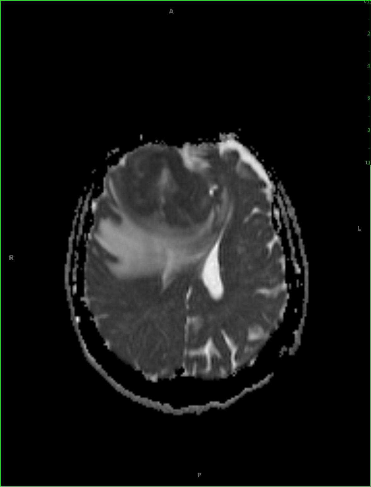


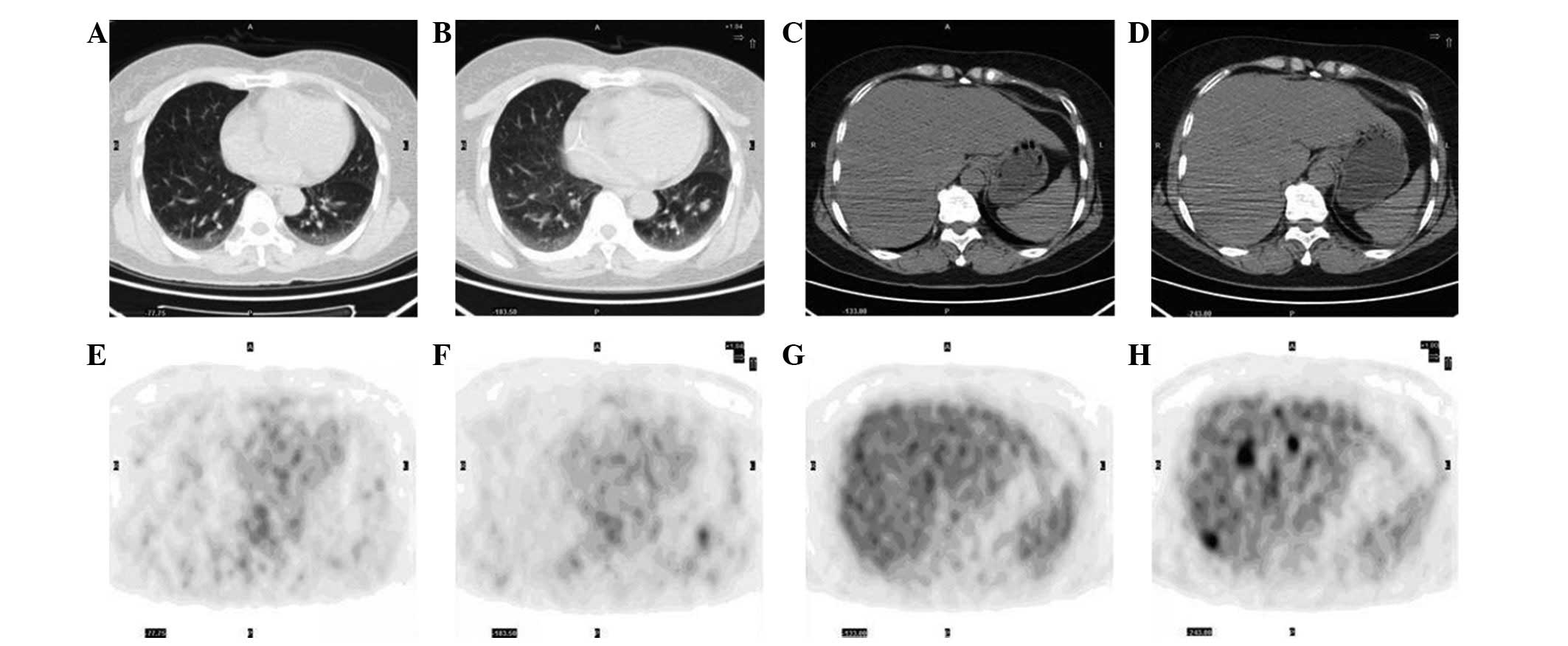
![The 4 stages of Lymphoma image by using PET/CT [17]. Focusing on ...](https://www.researchgate.net/publication/366208581/figure/fig4/AS:11431281106906315@1670896784740/The-4-stages-of-Lymphoma-image-by-using-PET-CT-17-Focusing-on-PET-MRI-scanners-since.png)


Create spaces with our architectural Cns Lymphoma Pet/ct gallery of comprehensive galleries of building images. spatially documenting photography, images, and pictures. perfect for architectural portfolios and presentations. The Cns Lymphoma Pet/ct collection maintains consistent quality standards across all images. Suitable for various applications including web design, social media, personal projects, and digital content creation All Cns Lymphoma Pet/ct images are available in high resolution with professional-grade quality, optimized for both digital and print applications, and include comprehensive metadata for easy organization and usage. Our Cns Lymphoma Pet/ct gallery offers diverse visual resources to bring your ideas to life. Reliable customer support ensures smooth experience throughout the Cns Lymphoma Pet/ct selection process. The Cns Lymphoma Pet/ct archive serves professionals, educators, and creatives across diverse industries. Comprehensive tagging systems facilitate quick discovery of relevant Cns Lymphoma Pet/ct content. Each image in our Cns Lymphoma Pet/ct gallery undergoes rigorous quality assessment before inclusion. Whether for commercial projects or personal use, our Cns Lymphoma Pet/ct collection delivers consistent excellence. Professional licensing options accommodate both commercial and educational usage requirements. Instant download capabilities enable immediate access to chosen Cns Lymphoma Pet/ct images. Our Cns Lymphoma Pet/ct database continuously expands with fresh, relevant content from skilled photographers.